

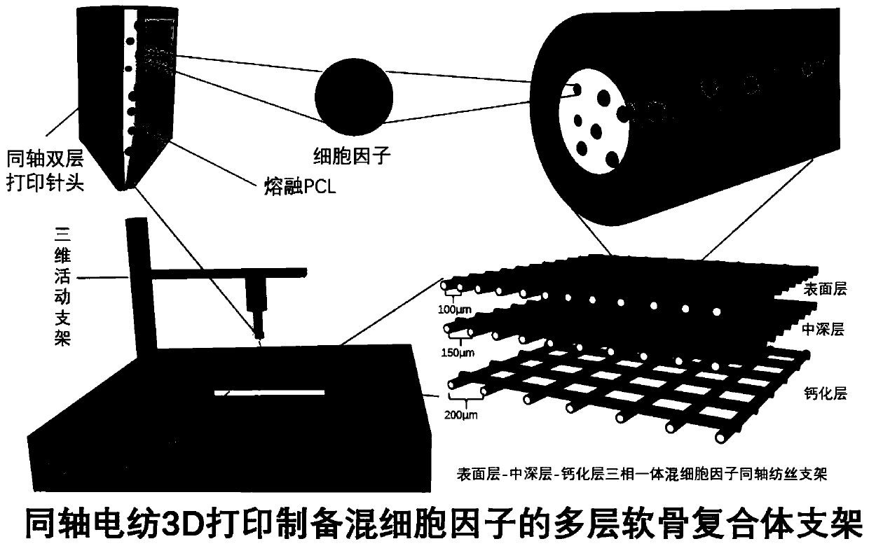
Preparation of coaxial electrospun-containing multilayered cartilage complex by electrospinning 3D printing
A coaxial electrospinning and 3D printing technology, applied in the field of 3D printing, can solve the problems that normal cartilage cannot provide friction resistance and support, and is easy to be damaged again, so as to promote cell proliferation and differentiation, improve printing accuracy, and high structural accuracy Effect
- Summary
- Abstract
- Description
- Claims
- Application Information
AI Technical Summary
Problems solved by technology
Method used
Image

Examples
preparation example Construction
[0033] S2, the preparation of three-phase integrated support;
[0034] S3, planting of cells, taking an appropriate amount of bone marrow mesenchymal stem cells to plant in each scaffold area.
[0035] Specifically, the method for culturing the seed cells in step S1 is that the bone marrow mesenchymal stem cells are cultured in a DMEM medium containing 10% fetal bovine serum in an incubator.
[0036] Specifically, the temperature in the incubator is 37°C, and there is 5% CO in the incubator 2 .
[0037] Specifically, the establishment of the model in the preparation step of the three-phase integrated stent in the step S2 includes the following steps;
[0038] S21, establishment of the model;
[0039] S22, preparation of materials;
[0040] S23. Equipment preparation.
[0041] Specifically, the specific process of establishing the model in the step S21 is: use three-dimensional printing software to establish a printing model, which is in the shape of a multi-layer cylinder...
Embodiment 1
[0047] (1) Use 3D printing software to build a printing model, which is multi-cylindrical, with different path gaps in the surface area, middle-deep area, and calcification layer area, and save it;
[0048](2) Measure an appropriate amount of polymer materials polycaprolactone and polylactic acid, and the polylactic acid is mixed with appropriate amounts of TGFβ1, BMP7, IGF cytokines and HA (hydroxyapatite) particles;
[0049] (3) Equipment preparation: adjust the process parameters of the electrospinning 3D printing equipment: the diameter of the printing wire is 10-50 μm, the printing temperature is 65°C, the air pressure of the barrel is 600-1000KPa during the printing process, and the voltage of the negative high voltage module is - 2.5~-50kV, the printing structure is controlled by the printing path, the printing path is 0 / 90°, the printing interval is 100μm, 150μm, 200μm, the polycaprolactone is loaded into the outer barrel, and the polylactic acid solution loaded with cy...
Embodiment 3
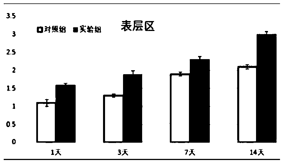
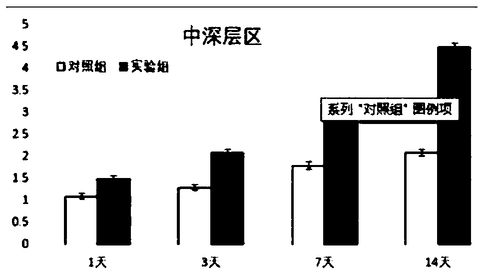
[0051] The biocompatibility of the obtained cartilage layered scaffold was characterized, and the obtained coaxial mixed cytokine three-phase integrated scaffold was planted with bone marrow stem cells and cultured in an incubator. The scaffold obtained by three-dimensional printing of pure polycaprolactone and bone marrow stem cells were used as the control group, and compared with each area of the three-phase scaffold. After cultured for 1, 3, 5, and 7 days, the cell viability in the surface area scaffold was detected by MTT method, which showed that the cells on the scaffold obtained by electrospinning 3D printing technology with cytokine microspheres had good proliferation behavior, and the cells The vitality is better, and the proliferation behavior is more significant than that of the control group.
PUM
| Property | Measurement | Unit |
|---|---|---|
| diameter | aaaaa | aaaaa |
Abstract
Description
Claims
Application Information