h based on electrochromic materials 2 s-activated probes and their biological applications
A technology of electrochromic materials and probes, applied in the direction of material excitation analysis, color/spectral characteristic measurement, wave energy or particle radiation treatment materials, etc., can solve the problems that have not been reported in the application, achieve fast response rate, high tissue penetration The effect of penetration depth and high activation multiple
- Summary
- Abstract
- Description
- Claims
- Application Information
AI Technical Summary
Problems solved by technology
Method used
Image
Examples
Embodiment 1
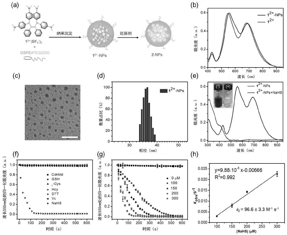
[0039] Embodiment 1. H based on electrochromic material 2 Construction of S-Activation Probes
[0040] 1. Synthesis of electrochromic materials.
[0041] Synthesize electrochromic material 1 according to the following route 2+ (EM 1 2+ ).
[0042]
[0043] 2. Encapsulated EM 1 2+ , to prepare nanoparticles 12+ -NPs.
[0044] In this example, in order to prepare 1 2+ -NPs, 0.43 mg 1 2+ (BF 4 - ) 2 and 10 mg of DSPE-PEG2000 were dissolved in 1 mL of tetrahydrofuran. Then inject this solution into 9 mL of secondary water rapidly during the sonication process, and continue to sonicate for 10 minutes. Next, tetrahydrofuran was removed at 65° C. by bubbling nitrogen. The obtained solution was ultrafiltered with a 10K ultrafiltration tube at 3500 rpm for 15 minutes to wash the sample, and the obtained solution was stored in a refrigerator at 4° C. until use. Depend on figure 2 As shown in (b), the nanoparticles 1 were successfully prepared 2+ -NPs; figure 2 Amon...
Embodiment 2
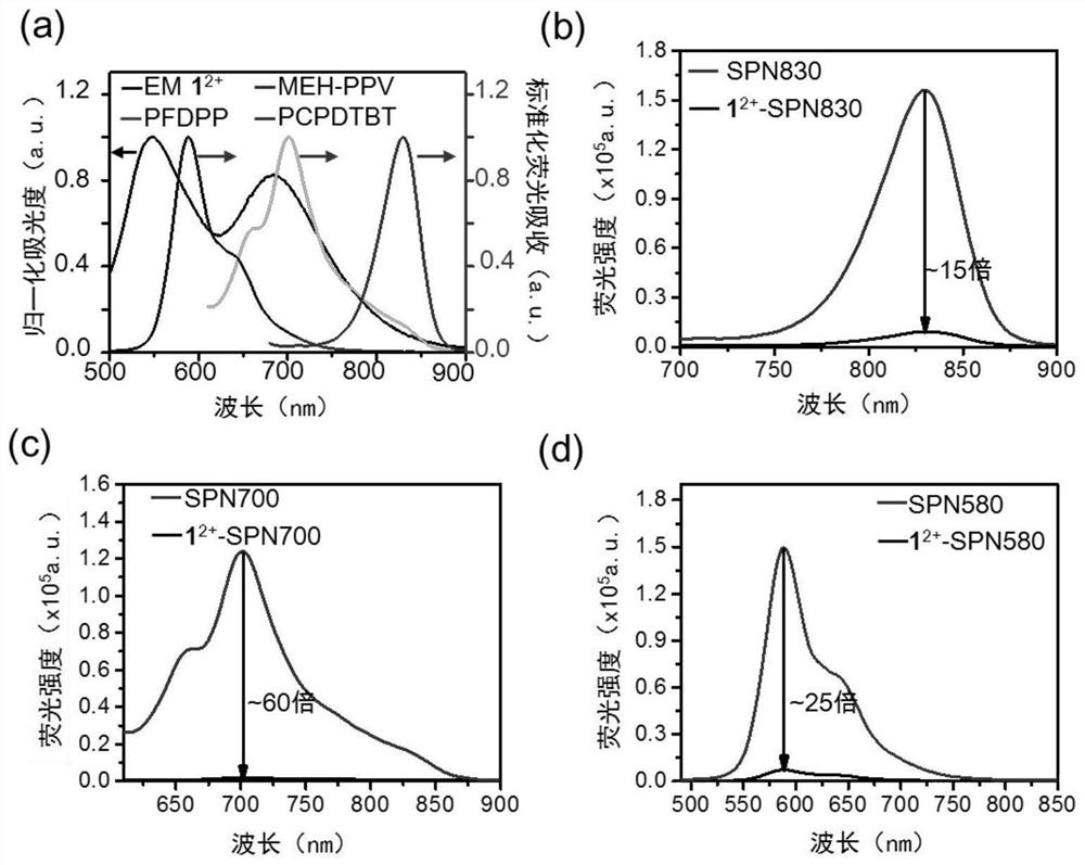
[0051] Embodiment 2. Probe 1 2+ -SNP830 on human serum H 2 S detection
[0052] This embodiment utilizes probe 1 2+ -SNP830 on human serum H 2 S was tested.
[0053] Specific method: using exogenous H 2 S was used as an internal standard for the determination of H in plasma 2 Concentration of S by adding 1 μL of ZnCl to 200 μL of human plasma 2 to remove H from plasma 2 S, then 1 μL of secondary water, 5, 10, 20, 30, 40 and 50 μM of HS were sequentially added to the other 6 groups of 200 μL of human plasma - , after that, add 1 2+ -SNP830 (final concentration is 28 μg / mL PCPDTBT, 48 μg / mL 1 2+ (BF 4 - ) 2 ), the treated sample was incubated at 37°C for 10 min, and the fluorescence spectrum of the solution was measured with a fluorometer, and the excitation wavelength was 650nm.
[0054] Figure 5 In, (a) and (b) both show probe 1 2+ -SNP830 can be detected by H in human serum 2 S activation, and can quantify H in human serum 2 The concentration of S was 12.7±3...
Embodiment 3
[0055] Example 3. Probe 1 2+ -SNP830 on live RAW264.7 intracellular H 2 S detection and in vivo imaging analysis
[0056] This embodiment utilizes probe 1 2+ -SNP830 on live RAW264.7 intracellular H 2 S was tested.
[0057] Specific method: RAW264.7 cells (~5×10 4 ) were inoculated in confocal culture dishes and cultured overnight. add 1 to the cell 2+ -SNP830 (28 / 48 μg / mL PCPDTBT / 1 2+ (BF 4 - ) 2 ), the probe was incubated at 37°C for 3 h, the medium was removed, the cells were washed once with cold PBS, NaHS (1 mM) was added for incubation for 1 h, and the cells were washed three times with cold PBS. In order to regulate the endogenous H 2 Production of S, added to RAW264.7 cells without any treatment L -Cys (0,0.1,0.2 and 0.5mM), then incubated for different times (0,0.5,1 and 2h). At the same time, in order to remove endogenous H 2 S, adding ZnCl to cells without any treatment 2 (0.3mM), the cells were incubated for 10min. In order to inhibit the activity o...
PUM
 Abstract
Description
Claims
Application Information
 - R&D
- Intellectual Property
- Life Sciences
- Materials
- Tech Scout
- Unparalleled Data Quality
- Higher Quality Content
- 60% Fewer Hallucinations
Browse by: Latest US Patents, China's latest patents, Technical Efficacy Thesaurus, Application Domain, Technology Topic, Popular Technical Reports.
© 2025 PatSnap. All rights reserved.Legal|Privacy policy|Modern Slavery Act Transparency Statement|Sitemap|About US| Contact US: help@patsnap.com


