A kind of porous bone repair scaffold material and preparation method thereof

A scaffold material and porous bone technology, applied in the field of biomedical composite materials, can solve the problems of poor mechanical properties of bone scaffold materials, affecting biocompatibility, insoluble in water, etc., and achieve high toughness, strength and good biocompatibility , Reduce the effect of the process link

Active Publication Date: 2020-07-03
NORTHWEST UNIV
View PDF7 Cites 1 Cited by
  • Summary
  • Abstract
  • Description
  • Claims
  • Application Information

AI Technical Summary

Problems solved by technology

However, animal collagen is not only difficult to dissolve in water, but also often has virus hidden dangers such as swine fever, mad cow disease, and rabies, so its use is limited
In addition, in the preparation of scaffold materials, intermolecular cross-linking of biomacromolecular collagen is required, and the selection of related cross-linking agents directly affects the physical, chemical and biological properties of the final prepared scaffold materials, such as chemical cross-linking The residual toxicity of the agent also directly affects the biocompatibility of the material. Due to the crosslinking performance of some enzyme crosslinking agents, the mechanical properties of the prepared bone scaffold material are poor, easy to swell, etc.

Method used


Image

  • A kind of porous bone repair scaffold material and preparation method thereof
  • A kind of porous bone repair scaffold material and preparation method thereof
  • A kind of porous bone repair scaffold material and preparation method thereof

Examples


Embodiment 1

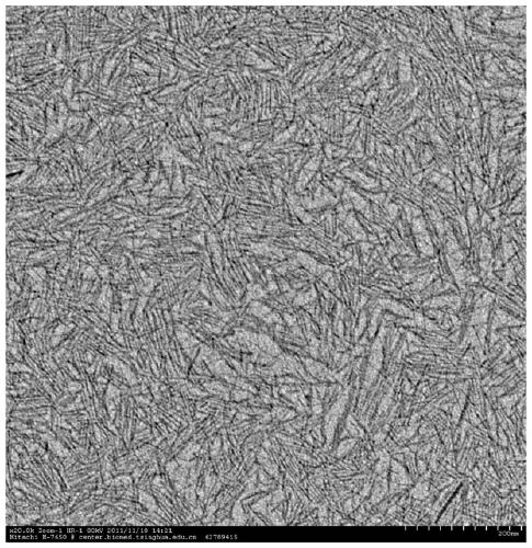
[0039]In a 100mL beaker, dissolve 5g of sodium oleate in 15mL of ethanol, and add 10mL of an aqueous solution containing 1g of human-like collagen to it, stir well, then add 10mL of 0.25M calcium chloride aqueous solution and 0.15M phosphoric acid 10 mL of sodium aqueous solution was stirred magnetically at room temperature (25° C.) for 2 days, the reaction solution was centrifuged to obtain a precipitate, and the precipitate was washed twice with ethanol to finally obtain sub-nanometer hydroxyapatite.

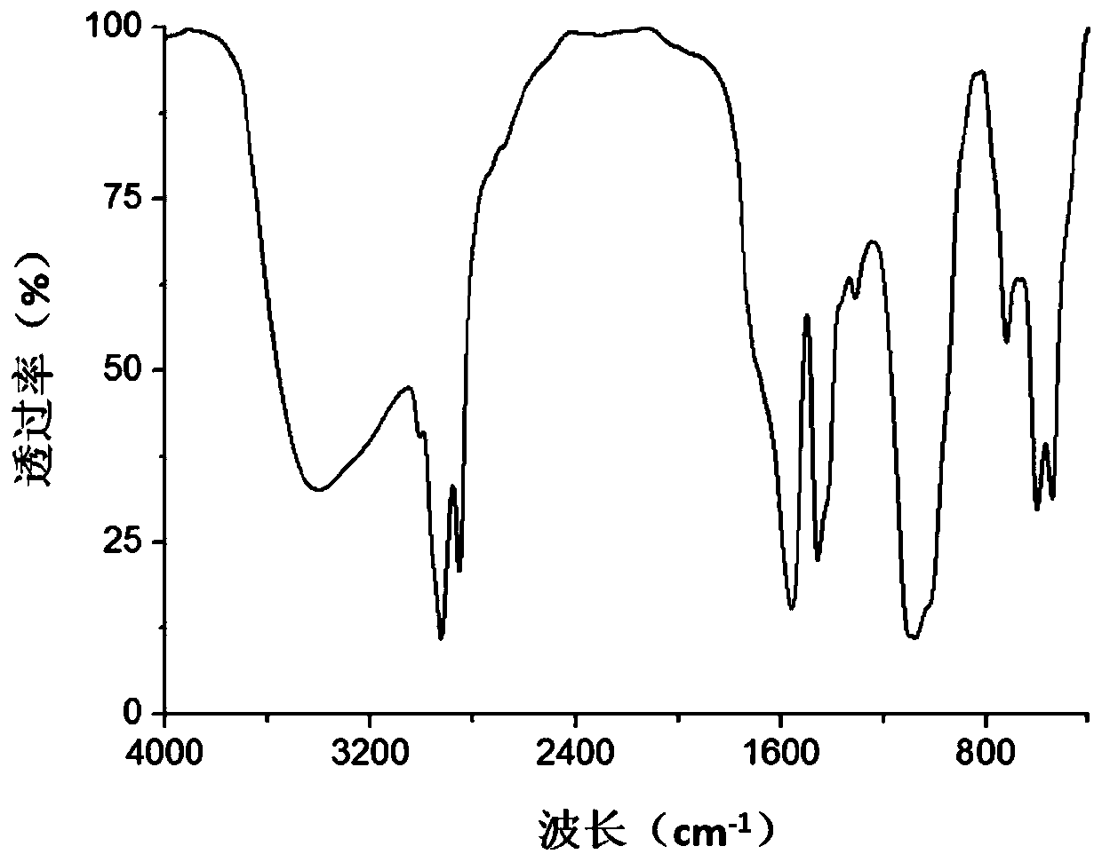
[0040] From figure 1 It can be seen from the figure that the sample prepared in this example is an ultrafine hydroxyapatite subnanowire with a diameter of about 1 nm and a length of about 40 nm to 100 nm; figure 2 It can be seen that the prepared samples are rich in hydroxyl groups and have similar characteristic absorption peaks with hydroxyapatite.

Embodiment 2

[0042] In a 100mL beaker, dissolve 5g of sodium oleate in 10mL of ethanol, and add 10mL of an aqueous solution containing 5g of human-like collagen to it, stir well, then add 10mL of 1.0M calcium chloride aqueous solution and 0.6M phosphoric acid 10 mL of sodium dihydrogen aqueous solution was magnetically stirred at room temperature (20° C.) for 5 days, the reaction solution was centrifuged to obtain a precipitate, and the precipitate was washed with ethanol 3 times to finally obtain sub-nanometer hydroxyapatite.

[0043] The physical and chemical characteristics of the sample prepared in this example are similar to those of the sample in Example 1, which are ultrafine hydroxyapatite subnanowires with a diameter of about 1 nm and a length of about 30 nm to 80 nm.

Embodiment 3

[0045] In a 100mL beaker, dissolve 5g of sodium oleate in 20mL of ethanol, and add 15mL of an aqueous solution containing 3g of human-like collagen to it, stir well, then add 15mL of 0.5M calcium chloride aqueous solution and 0.3M phosphoric acid After 15 mL of disodium hydrogen aqueous solution was magnetically stirred at room temperature (22°C) for 4 days, the reaction solution was centrifuged to obtain a precipitate, and the precipitate was washed with ethanol 3 times to finally obtain subnanometer hydroxyapatite.

[0046] The physical and chemical characteristics of the sample prepared in this example are similar to those of the sample in Example 1, which are ultrafine hydroxyapatite subnanowires with a diameter of about 1 nm and a length of about 30 nm to 100 nm.

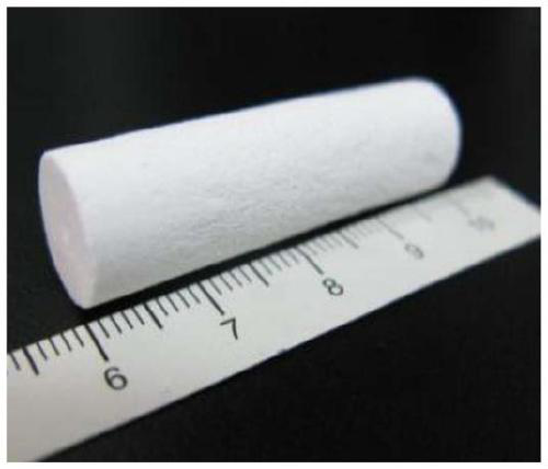
[0047] Preparation of porous bone repair scaffold materials:


PUM

PropertyMeasurementUnit
concentrationaaaaaaaaaa
pore sizeaaaaaaaaaa

Abstract

The invention discloses a porous bone repair scaffold material. The porous bone repair scaffold material is prepared by cross-linking reaction of human-like collagen and sub-nanometer hydroxyapatite under the action of a cross-linking agent. The cross-linking The agent is Agaricus bisporus polyphenol oxidase, the mass ratio of human-like collagen and sub-nanometer hydroxyapatite is 1: (2-5), and the dosage of Agaricus bisporus polyphenol oxidase is 400-1000U / g. In addition, the invention also discloses a preparation method of porous bone repair scaffold material. The invention uses Agaricus bisporus polyphenol oxidase as a cross-linking agent, sub-nanometer hydroxyapatite prepared by mineralization using human-like collagen as a template as an inorganic phase, human-like collagen as an organic phase, and added lecithin and Cell growth factor, the porous bone repair scaffold material prepared by enzymatic cross-linking technology does not require the removal of cross-linking agent residues, has good biocompatibility, and the scaffold material can be reused as a raw material for new bone formation after degradation.

Description

technical field [0001] The invention belongs to the technical field of biomedical composite materials, and in particular relates to a porous bone repair scaffold material and a preparation method thereof. Background technique [0002] Due to the increasing number of traffic accidents, bone injury diseases caused by sports trauma, and bone defects caused by bone necrosis, joint necrosis, trauma, infection, tumor resection, etc., plus in plastic surgery, maxillofacial repair and other operations, the The demand for bone scaffold materials is increasing day by day. As an implantable bone scaffold material, in order to facilitate the adhesion, proliferation and differentiation of cells, and to guide the growth of blood vessels, it has high biological activity and does not cause immune rejection of the body. It not only needs to have a three-dimensional penetrating microarchitecture and a suitable pore size, but also needs to have a microenvironment similar to that of natural bo...

Claims


Application Information

Patent Timeline
Patent Type & Authority Patents(China)
IPC IPC(8): A61L27/56A61L27/50A61L27/24A61L27/12A61L27/54A61L27/22C08J9/28C08J3/24C08L89/00
Inventor 惠俊峰贺其雅范代娣郑晓燕陈昱桥毛淳怡杨智健
Owner NORTHWEST UNIV
Why Eureka
Try Eureka