Method for identifying BPDE-added target protein

An identification method and target protein technology, applied in measuring devices, instruments, scientific instruments, etc., can solve problems such as lack, impossibility of chemical modification, discount of protein information, etc.

Pending Publication Date: 2019-08-16
SHANXI MEDICAL UNIV
3 Cites 4 Cited by

AI-Extracted Technical Summary

Problems solved by technology

[0004] 1. For the adducted target protein of BPDE, it is usually speculated through the change of content or function, and there is no direct evidence
[0005] 2. The identification method of other small molecular compound adducted target protein needs to chemically modify the small molecular compound, such as adding biological labels such as biotin or fluorescein. The modification process may change the activity of the small molecule
Compared with traditional methods, the adducted target protein screening technology based on this principle does not require any modification and labeling of small molecules, preserves the original characteristics of binding to the target protein and is independent of any biological effects of the drug, avoiding the need for any modification of t...

Abstract

The invention relates to the field of identification of BPDE-added target proteins, in particular to a method for identifying a BPDE-added target protein. The method comprises the steps of: 1) cell collection and protein extraction; 2) contamination and protease digestion of cell lysates; 3) mass spectrum pre-sample preparation; 4) LC-MS/MS analysis: taking peptide fragments to perform chromatographic separation, and performing DDA mass spectrometry by using a mass spectrometer after peptide fragment separation; and 5) mass spectrum database search: downloading a protein database, and obtaining a BPDE target protein according to mass spectrum analysis data. By adopting the method, the target protein which can be added with BPDE in the protein lysates can be directly identified, no chemicalmodification needs to be performed on micromolecules in a detection process, the original activity of the micromolecules is not affected, no influence is generated by any cell or tissue type, no requirement is proposed for pure protein, and even whole cell lysates can be used, so that the method can be widely applied to the identification of micromolecule-added target proteins.

Application Domain

Component separation

Technology Topic

Mass spectrum analysisChromatographic separation +18

Image

  • Method for identifying BPDE-added target protein
  • Method for identifying BPDE-added target protein
  • Method for identifying BPDE-added target protein

Examples

  • Experimental program(1)

Example Embodiment

[0040] Example:
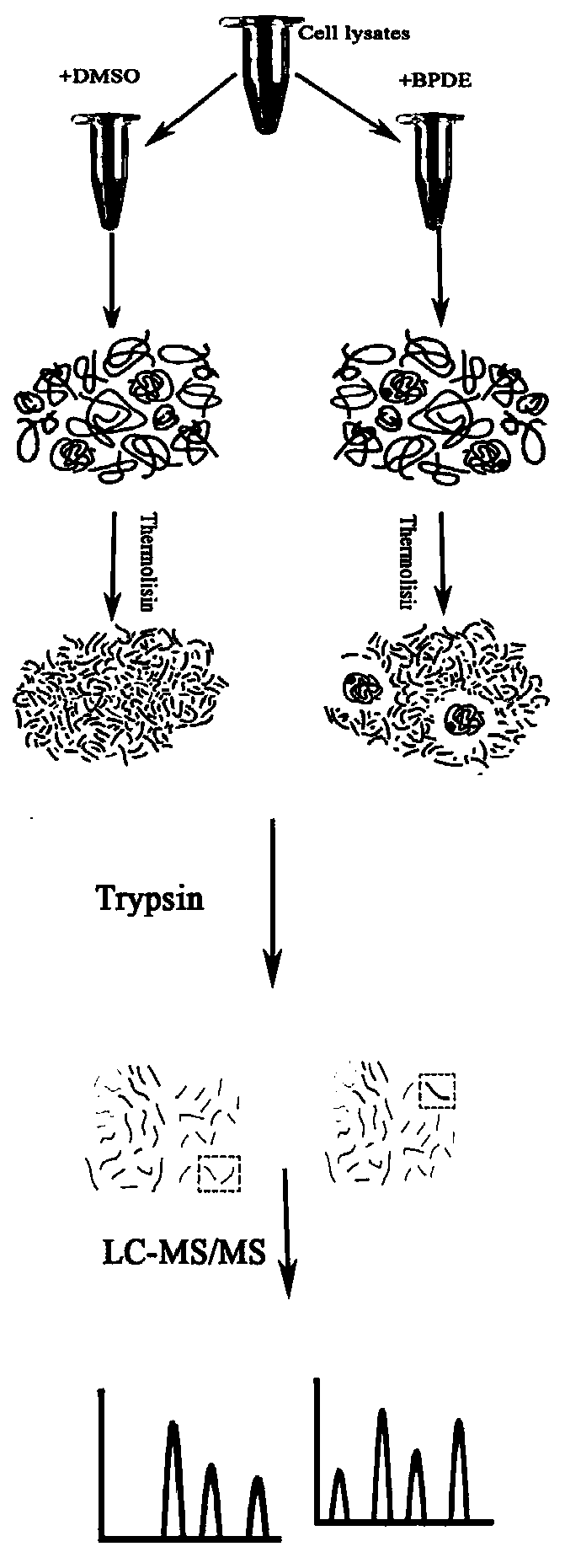
[0041] The identification method of BPDE adduct target protein in human neuroma blastoma cell (SH-SY5Y) lysate provided in the examples (see the method flow chart). figure 1 ) and results, including the following steps:
[0042] Step 1. SH-SY5Y cell culture and protein extraction
[0043] The neuroblastoma cell line SH-SY5Y was incubated at 37°C in 5% CO. 2 Cultivated under conditions, passaged (2-3 days/time), when the cell confluency reached 80% to 85%, poured off the medium, washed the cells twice with 5 mL of pre-cooled PBS buffer, and added trypsin to digest the cells. Centrifuge at 1000 rpm for 5 min, resuspend in 5 mL of pre-cooled PBS buffer, discard the supernatant after centrifugation (repeat the cycle to wash the cells 3 times), add 100 μL of pre-cooled PBS buffer to suspend the cells, and quickly freeze in liquid nitrogen for 1 min. Shake and thaw in a 25°C water bath (repeat this freeze-thaw cycle 4 times), centrifuge at 20,000 × g (gravity acceleration) and 4°C for 20 minutes, draw the supernatant and place it in a new centrifuge tube, discard the precipitate, i.e. for the extracted protein samples.
[0044] Step 2. Poisoning and protease digestion of cell lysates
[0045] Pipette part of the protein sample into a new centrifuge tube, use the Bradford method to determine the protein concentration in the sample, add 10 × TNC buffer (at the ratio of 9 (protein): 1 (TNC buffer)) and adjust the protein concentration to 6g /L (with pre-cooled PBS buffer), the lysate after adjusting the concentration was equally divided into two parts, respectively with 20mmol/L BPDE solution (the solvent is DMSO) and the same volume of solvent (DMSO), according to each 100μL Add 30 μL of BPDE solution to the lysate for moderate mixing (avoid vigorous shaking or vortexing during the mixing process), and incubate at room temperature for 2 h. After incubation, take 100 μL of each case, add thermolysin (according to the ratio of adding 1 μg thermolysin per 10 μg lysate), digest at 37 °C for 20 min, and add 3 μL of pre-cooled 50× protease inhibitor to each tube. agent, mix well and place on ice. In the present invention, any protease inhibitor capable of inhibiting thermolysin can be used, preferably a protease inhibitor capable of mass spectrometry, and 50× protease inhibitor produced by Biyuntian was used in the actual operation of this example.
[0046] Step 3. Sample preparation before mass spectrometry
[0047] To the samples treated with thermolysin, an appropriate amount of 8M urea lysate was added respectively. After low-temperature ultrasonic treatment, centrifuge at 4°C and 16,000 × g for 15 min, collect the supernatant, and use the Bradford method for protein quantification. Take 120 μg of each case. Carry out enzymatic hydrolysis (add dithiothreitol to 10mM (concentration of dithiothreitol) in each sample, 37°C, 2h, then cool to room temperature and add an appropriate amount of indoleacetic acid to 50mM (concentration of indoleacetic acid) , shake at 600rpm for 1min, dark at room temperature for 30min, add 100mM ammonium bicarbonate solution to dilute 6 times, add 4μg trypsin, shake at 600rpm for 1min, incubate at 37°C for 16-18h, add an appropriate amount of 1% trifluoroacetic acid solution to the final sample to stop the reaction .) The enzymatically hydrolyzed peptides were desalted using a C18 Cartridge solid phase extraction column, lyophilized in vacuum, reconstituted with 0.1% formic acid aqueous solution, and the peptide concentrations were determined for LC-MS analysis.
[0048] Step 4. LC-MS/MS analysis
[0049] An appropriate amount of peptides for each sample was chromatographically separated using an Easy nLC 1200 chromatographic system with a nanoliter flow rate. The buffer solution: A solution is 0.1% formic acid aqueous solution, B solution is 0.1% formic acid acetonitrile aqueous solution (acetonitrile is 85%). The column was equilibrated with 95% liquid A. The sample was injected into a Trap Column (100μm*20mm, 5μm, C18, Dr. Maisch GmbH) and then passed through a chromatographic analysis column (75μm*150mm, 3μm, C18, Dr. Maisch GmbH) for gradient separation with a flow rate of 300nL/min. The liquid phase gradient settings are as follows: 0min-2min, the linear gradient of liquid B is from 5%-8%; 2min-90min, the linear gradient of liquid B is from 8%-23%; 90min-100min, the linear gradient of liquid B is from 23%-40% ; 100min-108min, the linear gradient of B solution is from 40%-100%; 108min-120min, B solution is maintained at 100%.
[0050] After peptide separation, DDA (Data Dependent Acquisition) mass spectrometry was performed using a Q-Exactive Plus mass spectrometer (Thermo Scientific). Analysis time is 120, detection mode: positive ion, precursor ion scan range: 300-1800m/z, primary mass spectrometry resolution: 70,000@m/z 200, AGC target: 1e6, primary maximum IT: 50ms. The peptide mass spectrometry analysis was collected according to the following method: after each full scan (full scan), the MS2 scan was triggered to collect the 20 highest intensity precursor ions (MS2scan), the MS2 resolution: 17,500@m/z 200, AGC target: 1e5, Secondary Maximum IT: 50ms, MS2ActivationType: HCD, Isolation window: 1.6Th, Normalized collision energy: 27. NOTE: During the entire process of steps 3 and 4, the temperature should not be higher than 37 °C. Since the optimal reaction temperature of thermolysin is 70°C, it has considerable stability in the pH range of 5-9.5. If the temperature is too high, the digestibility of thermolysin is enhanced enough to completely digest all proteins in the lysate, resulting in failure of mass spectrometry identification.
[0051] Step 5. Search the mass spectrometry database
[0052] The mass spectrometry database search software used is MaxQuant1.6.0.16; the protein database used is from the uniprot Protein Database, the species is Homo Sapiens (human), and a total of 174301 protein sequences were downloaded on September 17, 2018. Among them, the MaxQuant library search software analysis parameter settings are shown in Table 1-1.
[0053] Table 1-1. MaxQuant software analysis parameter table
[0054]
[0055]
[0056] The protein samples containing the BPDE adduct target protein were compared with the protein samples without the BPDE adduct target protein, and the differential proteins between the two were screened. Through this identification method, a total of 428 BPDE adduct target proteins were identified. The specific results See Exhibit 1.
[0057]
[0058]
[0059]
[0060]
[0061]
[0062]
[0063]
[0064]
[0065]
[0066]
[0067]

PUM

no PUM

Description & Claims & Application Information

We can also present the details of the Description, Claims and Application information to help users get a comprehensive understanding of the technical details of the patent, such as background art, summary of invention, brief description of drawings, description of embodiments, and other original content. On the other hand, users can also determine the specific scope of protection of the technology through the list of claims; as well as understand the changes in the life cycle of the technology with the presentation of the patent timeline. Login to view more.
Why Eureka
Try Eureka