
Fluorescence immunochromatography combined detection kit and preparation method thereof

A detection kit and detection card technology, which is applied in the field of clinical medical examination, can solve problems such as low accuracy, complicated test operation, and long detection time, and achieve the effects of minimizing false positives, good repeatability, and easy automation

Inactive Publication Date: 2019-12-10
CHANGZHOU BIOWIN BIOPHARM
View PDF2 Cites 4 Cited by
  • Summary
  • Abstract
  • Description
  • Claims
  • Application Information

AI Technical Summary

Problems solved by technology

[0004] Chinese invention patent CN201010102136.5 discloses an ELISA kit containing vascular peroxidase protein and antibody. This method judges the severity of coronary heart disease by a single vascular peroxidase concentration, and the diagnostic accuracy of a single index is not high. , and the accuracy of ELISA method is not high, it is prone to false positives, and the detection time is long, and the test operation is complicated
Chinese invention patent CN201410623721.8 discloses a serum marker Samd3 protein ELISA kit for the diagnosis of coronary heart disease. The ELISA method is also used, but the accuracy is not high, false positives are prone to occur, and the detection takes a long time and the test operation is complicated. Accurate early diagnosis, risk prediction and severity assessment of serious cardiovascular events

Method used


Image

  • Fluorescence immunochromatography combined detection kit and preparation method thereof
  • Fluorescence immunochromatography combined detection kit and preparation method thereof
  • Fluorescence immunochromatography combined detection kit and preparation method thereof

Examples


Embodiment 1

[0033] (1) Preparation of sample pad

[0034] Use glass cellulose membrane as the sample pad, dissolve Nacl, BSA and other salt ions and proteins with buffer Tris, etc., then add a small amount of surfactant Tween20, adjust the pH to 7-8, and evenly spread it on the glass fiber according to the water absorption of the glass fiber , placed at 37 ° C and dried for 8 hours.

[0035] (2) Preparation of binding pads

[0036] Dilute the fluorescent microsphere-labeled monoclonal antibody with microsphere diluent, spread evenly on the treated sample pad, seal it after vacuum freeze-drying, and store it at room temperature. The preparation process of the fluorescent microsphere-labeled antibody is as follows:

[0037] a. Coupling of fluorescent microspheres with anti-high-sensitivity C-reactive protein monoclonal antibody

[0038]Fluorescent microspheres with a particle size of 300nm were first activated with EDC and NHS activators and stabilizers, washed after activation to remove ...

Embodiment 2

[0051] (1) Preparation of sample pad

[0052] Use glass cellulose membrane as the sample pad, dissolve Nacl, BSA and other salt ions and protein with buffer PBS, etc., then add a small amount of surfactant TritonX-100, adjust the pH to 7-8, spread evenly on the glass according to the water absorption of the glass fiber. On the fiber, dry at 37°C for 8 hours.

[0053] (2) Preparation of binding pads

[0054] Dilute the fluorescent microsphere-labeled monoclonal antibody with microsphere diluent, spread evenly on the treated sample pad, seal it after vacuum freeze-drying, and store it at room temperature. The preparation process of the fluorescent microsphere-labeled antibody is as follows:

[0055] a. Coupling of fluorescent microspheres with anti-high-sensitivity C-reactive protein monoclonal antibody

[0056] Fluorescent microspheres with a particle size of 300nm were first activated with EDC and NHS activators and stabilizers, washed after activation to remove the activato...

Embodiment 3

[0069] Detection of hypersensitive C-reactive protein, myeloperoxidase and lipoprotein phospholipase A2 fluorescent immunochromatographic detection kit.

[0070] (1) Draw a standard curve

[0071] Add different concentrations of hsCRP, myeloperoxidase, and lipoprotein phospholipase A2 antigen standards on the kit sample pad prepared according to Example 1 (7 different concentrations are taken respectively, hsCRP antigen standard Standard products were 0, 0.5, 5, 10, 40, 100, 150ng / mL, myeloperoxidase antigen standard products were 0, 10, 50, 100, 200, 500, 1000ng / mL, lipoprotein phospholipase A2 antigen The standard products were 0, 50, 100, 200, 500, 1000 and 1500ng / mL respectively, and each concentration was set in 3 repetitions), after 15 minutes, placed in a fluorescence quantitative immunochromatography instrument to obtain the fluorescence signal intensity, according to the detection line Calculate the concentration of the tested sample from the standard curve based on ...


PUM

PropertyMeasurementUnit
Particle sizeaaaaaaaaaa
Emission wavelengthaaaaaaaaaa

Abstract

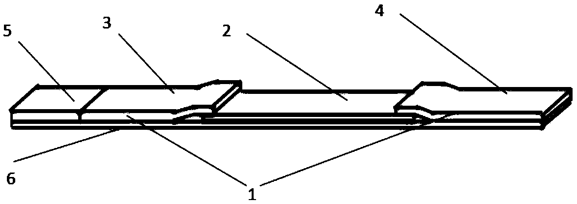
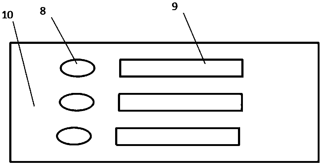
The invention discloses a preparation method of a composite fluorescence immunochromatography detection kit for quantitatively detecting the hypersensitive C-reactive protein (Hs-CRP), myeloperoxidase(MPO) and lipoprotein phospholipase A2 (Lp-PLA2). The detection kit comprises a drying agent, a sealing bag and a detection card, the detection card comprises a triplet buckle and a reagent strip, and the reagent strip comprises a sample pad, a combination pad, a blood filtering membrane, a nitrocellulose membrane, an absorbent paper and a PVC bottom plate. The combination pad is coated with an hsCRP antibody, an MPO antibody and an Lp-PLA2 antibody, the hsCRP antibody is one or a combination of an hsCRP monoclonal antibody and an hsCRP polyclonal antibody, the MPO antibody is one or a combination of an MPO monoclonal antibody and an MPO polyclonal antibody, and the Lp-PLA2 antibody is one or a combination of an Lp-PLA2 monoclonal antibody and an Lp-PLA2 polyclonal antibody. The method disclosed by the invention has the outstanding advantages of specificity, sensitivity, high yield, rapidness, simplicity, convenience, good repeatability, easiness in automation, minimized false positive, high detection efficiency, low detection cost and the like, and has the important practical significance.

Description

technical field [0001] The invention relates to the field of clinical medical examination, in particular to a fluorescent immunochromatographic detection kit for combined quantitative detection of hypersensitive C-reactive protein, myeloperoxidase and lipoprotein phospholipase A2 and a preparation method thereof. Background technique [0002] Cardiovascular disease is a common clinical emergency and severe disease, with a high fatality rate and a dangerous condition, which seriously threatens human health and life. According to data, the basic pathological change of cardiovascular disease is atherosclerosis. Recent studies have shown that inflammation is the basis of atherosclerosis and cardiovascular diseases. In particular, chronic inflammation is considered to increase the risk of arterial damage and disease. Atherosclerotic plaques are prone to rupture and play a key role in the formation of atherosclerosis. Arterial damage is the result of a chronic inflammatory respon...

Claims


Application Information

Patent Timeline
IPC IPC(8): G01N33/68G01N33/533G01N33/58G01N33/558
CPCG01N33/533G01N33/558G01N33/582G01N33/6803G01N2496/05
Inventor 章燕李亚楠刘凤鸣
Owner CHANGZHOU BIOWIN BIOPHARM
Learn More