Magnetic nanoparticle for imaging guidance and preparation method thereof

A technology of magnetic nanoparticles and alloy nanoparticles, which is applied in the field of biomedicine, can solve the problem of high relaxation rate of spherical particles and other issues, and achieves the effect of increasing stability and not easy to fall off.

Active Publication Date: 2019-12-13
NANJING XIAOZHUANG UNIV
View PDF12 Cites 3 Cited by
  • Summary
  • Abstract
  • Description
  • Claims
  • Application Information

AI Technical Summary

Problems solved by technology

As stated in the specification, when Fe 3 o 4 Polymerization of polymer materials on the surface of magnetic nanoparticles can improve the biocompatibility of nanoparticles, but whether the final spherical particles meet the high relaxation rate remains to be discussed.

Method used


Image

  • Magnetic nanoparticle for imaging guidance and preparation method thereof
  • Magnetic nanoparticle for imaging guidance and preparation method thereof
  • Magnetic nanoparticle for imaging guidance and preparation method thereof

Examples


preparation example 1Fe3

[0047] Preparation Example 1Fe 3 o 4 Preparation of nanoparticles

[0048] FeSO 4 ·7H 2 O 4.2g and FeCl 3 ·6H 2 O 5.4g were respectively dissolved in 50mL water, shaken to fully dissolve the crystals, put the two solutions into the three-necked flask, shake and shake well, add ammonia water with a mass concentration of 30% in the dropping funnel, and fill the three-necked flask with nitrogen for 2 Minutes, under the protection of nitrogen, add ammonia water drop by drop to the reaction system until the pH of the system is about 12.0, stir vigorously in a water bath at 55°C for 50 minutes at a constant temperature, centrifuge after the reaction, remove the supernatant, and wash the precipitate repeatedly with distilled water To neutrality, pour off the supernatant, dry in vacuum at 65°C, and grind to obtain Fe 3 o 4 Nanoparticles.

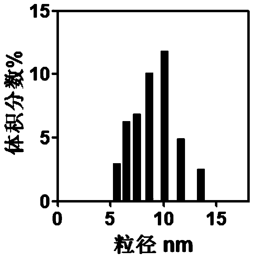
[0049] For the prepared Fe 3 o 4 Nanoparticles were characterized, figure 1 Indicates Fe 3 o 4 The particle size distribution diagram ...

preparation example 2

[0050] Preparation example 2 Fe coated with mesoporous silica on the surface 3 o 4 Preparation of nanoparticles

[0051] The Fe that Preparation Example 1 obtains 3 o 4 Disperse 1mmol of nanoparticles in 100mL of 50°C aqueous solution, adjust the pH value to 9.0, add 10mL of tetraethyl orthosilicate and 25mL of ethyl acetate under stirring, stir for 10min to dissolve, then add 3-aminopropyl-triethoxy Silane 1.5mL, keep the temperature of the reaction solution at 65°C, stir the reaction for 24 hours, cool the product, centrifuge, and wash with ethanol 6 times to obtain the Fe powder coated with mesoporous silica. 3 o 4 Nanoparticles, dispersed in water for later use.

preparation example 3

[0052] Preparation Example 3 Preparation of gold nanoparticles

[0053] Lipoic acid was dissolved in deionized water to prepare a solution with a concentration of 0.1 mM, and HAuCl 4 ·3H 2 O crystals were dissolved in deionized water to prepare a solution with a concentration of 1 mM; the NaBH 4 Dissolve in deionized water to prepare a solution with a concentration of 0.1M. Take a glass vial and add 4mL ddH 2 After O, deoxygenate with nitrogen for 2min, then add lipoic acid aqueous solution (0.5mL, 0.1mM) and HAuCl 4 Aqueous solution (0.6mL, 1mM), after incubating at 37°C for 10min under ultrasonic conditions, add NaBH 4 solution (15 μL, 0.1 M), after ultrasonication for 20 min until the color of the solution changed from light yellow to purple, and stored at 4°C until use.

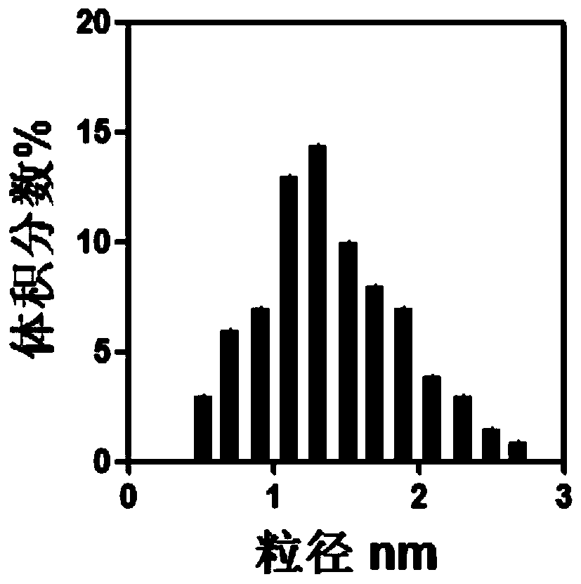
[0054] The prepared gold nanoparticles were characterized, and the results were as follows: figure 2 as shown, figure 2 It is the particle size distribution diagram of gold nanoparticles. It can ...


PUM

PropertyMeasurementUnit
Particle sizeaaaaaaaaaa

Abstract

The invention discloses a magnetic nanoparticle. The magnetic nanoparticle takes Fe3O4 nanoparticles as a core, the surface of the magnetic nanoparticle is coated with mesoporous silica, the surface of the mesoporous silica material is modified with a photosensitizer and embedded alloy nanoparticles, and the photosensitizer is selected from one of hematoporphyrin, hematoporphyrin monomethyl ether,chlorin and 5-aminolevulinic acid. The magnetic nanoparticle prepared by the invention can be used as a nuclear magnetic resonance contrast agent to carry out imaging positioning on tumors, and thennuclear magnetic resonance imaging is used as guidance to carry out photodynamic therapy on the tumors.

Description

technical field [0001] The invention belongs to the technical field of biomedicine, and in particular relates to a magnetic nanoparticle for magnetic resonance imaging to guide the treatment of brain tumors, a preparation method and application thereof. Background technique [0002] Tumor is the first major disease that threatens human life and health today. Among them, brain tumors are the most difficult to overcome because of their special growth sites. With the development of social life, the electrical appliances that people use daily, in addition to conventional TVs and refrigerators, computers, mobile phones, etc. have become indispensable tools for people's work and life. However, these electrical appliances have electromagnetic radiation, which can induce brain tumors. happened. In some cases, women are often exposed to computer radiation during pregnancy, which affects the normal growth and development of the fetus after long-term exposure, resulting in congenital ...

Claims


Application Information

Patent Timeline
IPC IPC(8): A61K49/08A61K49/18B82Y5/00B82Y15/00A61K41/00A61K47/04A61K47/02A61K47/52A61P35/00
CPCA61K41/0061A61K41/0071A61K47/02A61K49/08A61K49/183A61K47/52A61P35/00B82Y5/00B82Y15/00
Inventor 唐宁张边江陈全战
Owner NANJING XIAOZHUANG UNIV
Why Eureka
Try Eureka