Bone cartilage repairing bracket and preparation method thereof

A technology of osteochondral and cartilage, applied in the field of osteochondral repair scaffold and its preparation, can solve the problems of cartilage tissue easily surpassing the surrounding normal tissue, growing into the scaffold, cartilage hyperplasia, etc., and achieve the effect of avoiding cartilage hyperplasia

Inactive Publication Date: 2020-01-07
SHENZHEN LANDO BIOMATERIALS
View PDF9 Cites 2 Cited by
  • Summary
  • Abstract
  • Description
  • Claims
  • Application Information

AI Technical Summary

Problems solved by technology

For example, after implantation in the body, the cartilage layer is in contact with other tissues and cells, and the external fibroblasts will grow into the cartilage layer through the pores of the cartilage layer or even grow into the scaffold, thereby affecting the normal bone structure. The regeneration process affects healing; at the same time, the repaired cartilage tissue tends to exceed the surrounding normal tissue, resulting in cartilage hyperplasia

Method used


Image

  • Bone cartilage repairing bracket and preparation method thereof
  • Bone cartilage repairing bracket and preparation method thereof
  • Bone cartilage repairing bracket and preparation method thereof

Examples


preparation example Construction

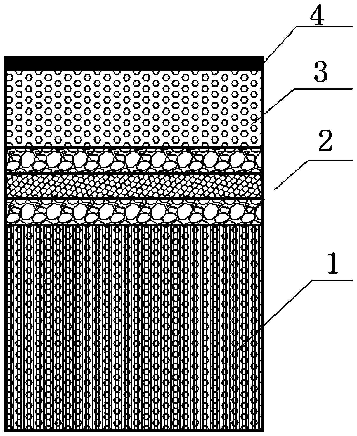
[0079] The embodiment of the present invention also discloses a method for preparing an osteochondral repair scaffold, which includes the following steps:

[0080] S10: preparing the subchondral bone layer, the calcified cartilage layer and the cartilage layer connected in sequence;

[0081] S20: forming a dense perichondrial layer on the surface of the cartilage layer away from the calcified cartilage layer.

[0082] In step S10, for the preparation of the subchondral bone layer, the calcified cartilage layer, and the cartilage layer that are sequentially connected, different preparation methods and different orders can be used.

[0083] In an embodiment of the present invention, first, polylactic acid (PLLA) is used to form a calcified cartilage layer by thermally induced phase separation. Then, a subchondral bone layer and a cartilage layer are formed on the surfaces of both sides of the calcified cartilage layer.

[0084] In this embodiment, in the thermally induced phase separatio...

Embodiment 1

[0106] (1) Preparation of calcified cartilage layer

[0107] Prepare a PLLA solution with a concentration of 4% (g / 100ml) (PLLA viscosity average molecular weight is 100000), the solvent is tetrahydrofuran, and pour it into a mold with a layer of fructose microsphere porogen (porogen particle size 100μm-200μm) at the bottom In the process, immerse the porogen, and form a layer of PLLA solution on the porogen; then add a layer of porogen on the top of the PLLA solution layer (the particle size of the porogen is 100μm-200μm), and then add PLLA The solution is filled with the porogen, and then thermally induced phase separation at -20°C for 2 hours, and then the solvent is replaced with pure water three times, then lyophilized to obtain a calcified cartilage layer, and the calcified cartilage layer after lyophilization is placed in a UV / ozone machine Treat for 10min to enhance its hydrophilicity.

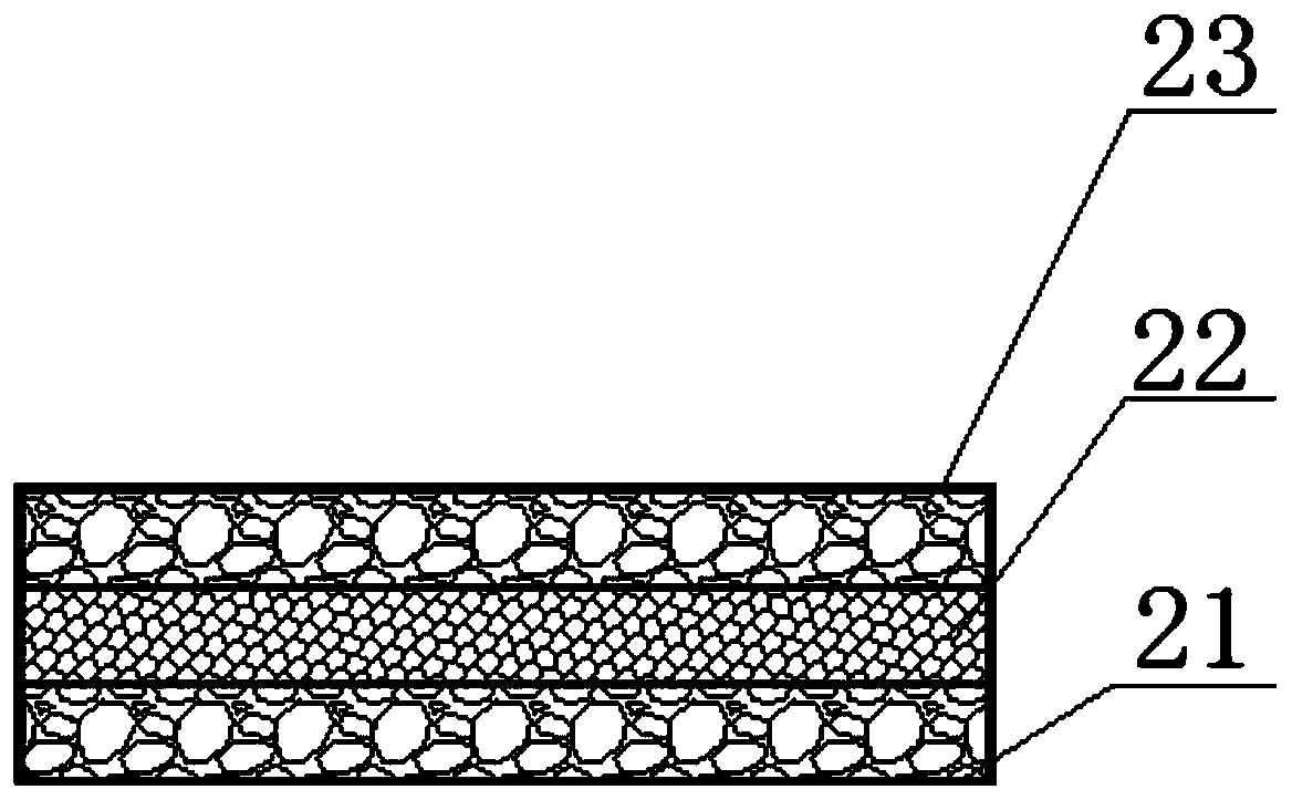
[0108] The calcified cartilage layer has two surface layers and a barrier layer locate...

Embodiment 2

[0122] (1) Preparation of calcified cartilage layer

[0123] In this embodiment, the preparation of the calcified cartilage layer is basically the same as that of Embodiment 1, except that in this embodiment, the thermally induced phase separation is performed at a temperature of -80° C. for 1 hour.

[0124] The resulting calcified cartilage layer has two surface layers and a barrier layer located between the two surface layers. In this embodiment, the PLLA solution immersed the porogen part to form two surface layers, the macropore diameter is 100-500μm, and the micropore diameter is 500nm-5μm; the PLLA solution layer is formed as a barrier layer, the pore size is 500nm-5μm .

[0125] (2) Preparation of subchondral bone layer

[0126] Preparation of metal frame

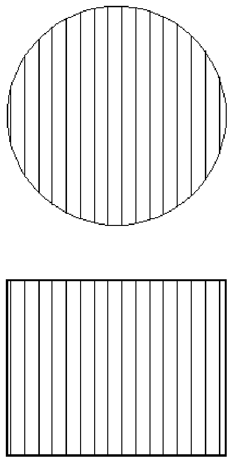
[0127] Use software to design the subchondral bone metal frame and print it with a three-dimensional printer. The material of the metal frame in this embodiment is magnesium alloy. The outer diameter of the metal frame is 10...


PUM

PropertyMeasurementUnit
particle diameteraaaaaaaaaa
pore sizeaaaaaaaaaa
pore sizeaaaaaaaaaa

Abstract

The invention relates to a bone cartilage repairing bracket. The bone cartilage repairing bracket comprises a cartilage lower bone layer, a calcification cartilage layer, a cartilage layer and a cartilage film layer which are sequentially arranged, wherein the cartilage film layer is a compact film layer away from one side surface of the calcification cartilage layer, of the cartilage layer; and the compact film layer can prevent the situation that external fiber cells grow into the cartilage layer. The invention further discloses a preparation method of the bone cartilage repairing bracket. According to the bone cartilage repairing bracket disclosed by the invention, the compact film layer is arranged on one side surface of the cartilage layer, and is used as a screen film of the cartilage layer, so that the condition that the external fiber cells grow into the cartilage layer can be avoided, the repaired cartilage tissue cannot exceed surrounding normal tissue, and cartilage proliferation is avoided.

Description

Technical field [0001] The invention relates to the technical field of tissue engineering, in particular to an osteochondral repair scaffold and a preparation method thereof. Background technique [0002] Cartilage injury is a kind of osteoarthritis, which is a common and multiple disease in the middle-aged and elderly people. The current clinical treatment methods cannot provide a good repair effect for the large-scale cartilage defects formed in the middle and late stages of osteoarthritis. Tissue engineering osteochondral repair scaffold is the most promising repair method. After the osteochondral repair scaffold is implanted in the body, it is gradually replaced with normal tissue in the body to complete the repair. In order to simulate the structure of natural osteochondral structure, the osteochondral repair scaffold is usually designed as a three-layer structure: cartilage layer, calcified cartilage layer and subchondral bone layer. [0003] The traditional three-layer str...

Claims


Application Information

Patent Timeline
Patent Type & Authority Applications(China)
IPC IPC(8): A61F2/30A61L27/40A61L27/24A61L27/18A61L27/12A61L27/02A61L27/50A61L27/56
CPCA61F2/30756A61F2/30942A61L27/24A61L27/18A61L27/12A61L27/025A61L27/56A61L27/50A61F2002/30766A61L2430/06C08L67/04
Inventor 潘洋魏欣苗陈雪霓谭荣伟佘振定
Owner SHENZHEN LANDO BIOMATERIALS
Why Eureka
Try Eureka