Mesoporous silica-based nano-drug delivery system capable of regulating drug release as well as construction and application of nano-drug delivery system
A technology of mesoporous silica and nano-drugs is applied in the fields of drug delivery, drug combination, and inactive medical preparations, etc. It can solve the problems of difficult to achieve precise drug release, complicated preparation of drug delivery systems, and long time-consuming.
- Summary
- Abstract
- Description
- Claims
- Application Information
AI Technical Summary
Problems solved by technology
Method used
Image
Examples
Embodiment 1
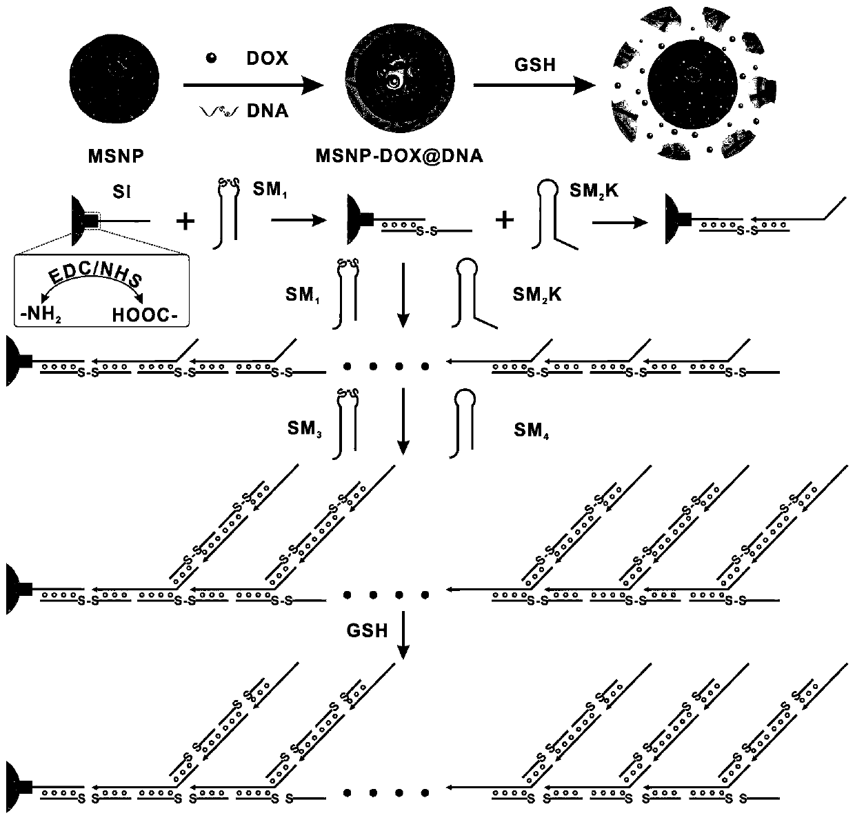
[0084] (1) 7μM L -1 SI was added to EDC and NHS at a molar ratio of 1:1 into PBS at pH=7.4, and incubated at 37°C for 20 minutes to obtain carboxyl-activated SI.
[0085] (2) Mix the above-mentioned activated SI with 0.5mgmL -1 The aminated MSNP was shaken and incubated at 37°C for 6h to obtain MSNP-SI.
[0086] (3) Mix the MSNP-SI obtained above with 8 μM L -1 SM 1 , 8μM L -1 SM 2 K, incubated at 37°C for 4h to obtain linearly polymerized MSNPs.
[0087] (4) Mix the above-mentioned linear polymerized MSNP with 8 μM L -1 SM 3 , 8μM L -1 SM 4 , and incubated at 37° C. for 10 h to prepare DNA-wrapped MSNP (MSNP@DNA).
Embodiment 2
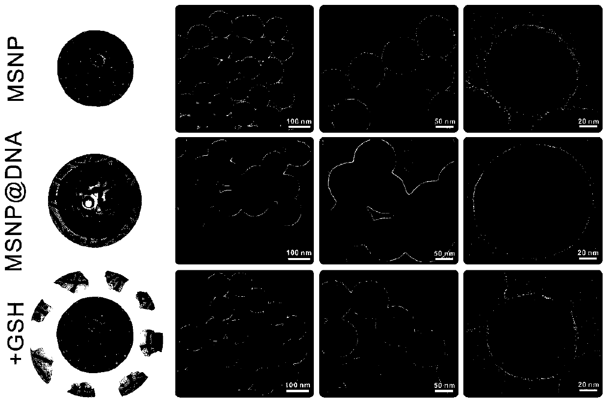
[0089] The nano-drug delivery system based on mesoporous silica constructed in Experimental Example 1 of the present invention was characterized by transmission electron microscopy.
[0090] (1) Take 10 μL of the MSNP@DNA sample prepared in Example 1 and add it dropwise to the carbon support film copper grid, let it stand for 10 minutes, absorb the solution with filter paper, and dry the copper grid with an infrared lamp.
[0091] (2) Observing the sample prepared above with a transmission electron microscope to observe its morphology. Such as figure 2 As shown, before unwrapping, MSNP has a porous structure and can load small molecule drugs; after DNA wrapping, the surface of MSNP has a shell layer with a thickness of ~10nm, indicating that DNA has successfully wrapped MSNP. Using 10mM glutathione (GSH) to incubate MSNP@DNA for 5h, it was found that the surface shell of MSNP disappeared, which indicated that GSH could break the disulfide bond and cleave the DNA shell. This...
Embodiment 3
[0093] In this experiment, ultraviolet-visible light spectroscopy was used to evaluate the loading of MSNP on chemotherapeutic drug DOX.
[0094] (1) drug load
[0095] 10mL0.5 mgmL -1 MSNP with 20 μM L -1 DOX was shaken and incubated at 37°C for 10 h, centrifuged and washed three times to obtain DOX-loaded MSNPs.
[0096] (2) Ultraviolet-visible light spectrum measurement
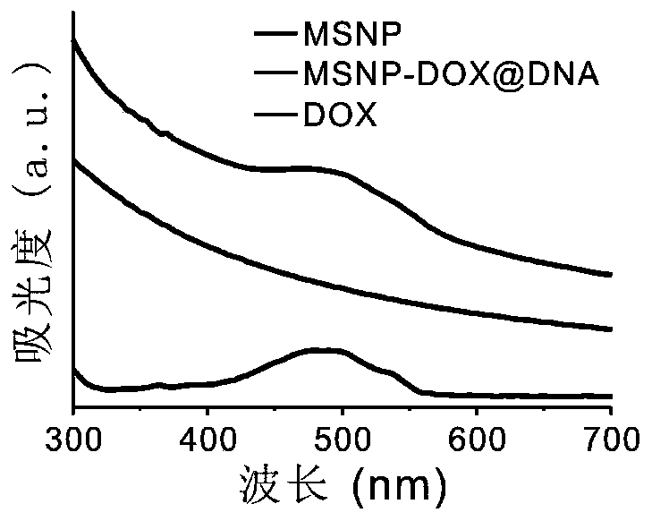
[0097] MSNP, DOX and MSNP-DOX@DNA were scanned in the range of 300-700nm by UV-visible spectroscopy, and their absorption characteristic peaks were observed.
[0098] (3) Results
[0099] Such as image 3 As shown, MSNP-DOX@DNA and DOX have the characteristic peak of DOX at 485nm, indicating that MSNP is successfully loaded with the chemotherapy drug DOX.
PUM
| Property | Measurement | Unit |
|---|---|---|
| thickness | aaaaa | aaaaa |
Abstract
Description
Claims
Application Information
 - R&D
- Intellectual Property
- Life Sciences
- Materials
- Tech Scout
- Unparalleled Data Quality
- Higher Quality Content
- 60% Fewer Hallucinations
Browse by: Latest US Patents, China's latest patents, Technical Efficacy Thesaurus, Application Domain, Technology Topic, Popular Technical Reports.
© 2025 PatSnap. All rights reserved.Legal|Privacy policy|Modern Slavery Act Transparency Statement|Sitemap|About US| Contact US: help@patsnap.com


