
Hog cholera virus envelope protein oligomeric protein and preparation method and application thereof

A swine fever virus and envelope protein technology, applied in the direction of viruses/phages, biochemical equipment and methods, viruses, etc., can solve the problem of incomplete understanding of the natural occurrence and mutation mechanism of the virus, and it is difficult to achieve modern prevention and control and eradication of swine fever Epidemic disease, the uncontrollable future trend of the virus, etc., to achieve the effect of easy large-scale production and purification, obvious protection effect, and good practical application value

Active Publication Date: 2020-02-28
许雁
View PDF3 Cites 3 Cited by
  • Summary
  • Abstract
  • Description
  • Claims
  • Application Information

AI Technical Summary

Problems solved by technology

For example, nucleic acid vaccines are slow in inducing antibodies, and the inoculation dose is large, which has the potential risk of integration and transformation of chromosomes; for humans, live virus vector vaccines cannot control this type of virus because they do not fully understand the natural occurrence and mutation mechanism of the virus. The future trend of the future; the subunit vaccine immunogenicity is usually worse than the virus particle antigen, and cannot provide effective protection; the gene deletion vaccine is usually a live attenuated vaccine, and its long-term use may change the original ecological environment and virus community of CSFV. Make the swine fever epidemic strain evolve away from the vaccine virus, and it is difficult to meet the requirements of modern prevention and control and eradication of swine fever

Method used


Image

  • Hog cholera virus envelope protein oligomeric protein and preparation method and application thereof
  • Hog cholera virus envelope protein oligomeric protein and preparation method and application thereof
  • Hog cholera virus envelope protein oligomeric protein and preparation method and application thereof

Examples


Embodiment 1

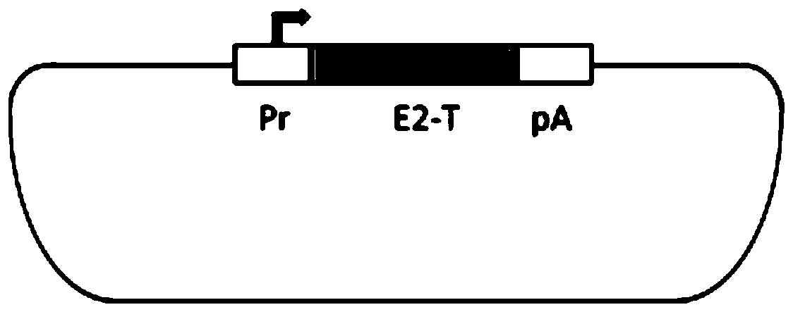
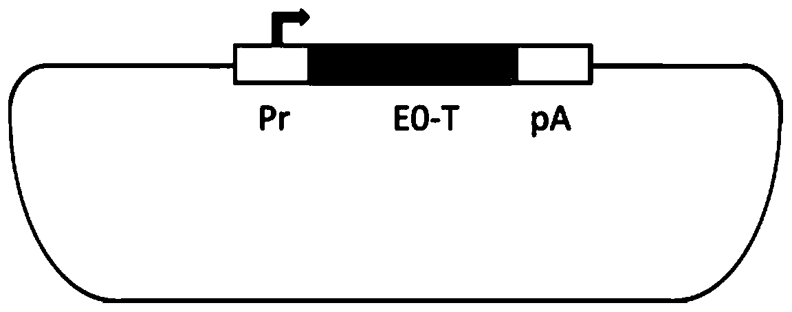
[0043] This example provides the construction and detection of the expression vectors of the envelope protein E2 oligomeric protein body and E0 oligomeric protein body of classical swine fever virus.

[0044] 1. Artificially synthesized DNA fragments of a single fusion subunit of an oligomeric protein body

[0045] In this example, the envelope protein E2 oligomeric protein body of swine fever virus contains four specially constructed envelope protein E2 fusion subunits, and each fusion subunit is composed of four parts: E2 subunit polypeptide, G6S9 short peptide link, western Nile virus C protein oligomerization structure fragment (WNV-Alpha4) and histidine (His) tag short peptide. Similarly, the envelope protein E0 oligomeric protein body of classical swine fever virus also contains four specially constructed envelope protein E0 fusion subunits, and each fusion subunit is composed of four parts: E0 subunit polypeptide, G6S9 short peptide link, western Nile virus C protein o...

Embodiment 2

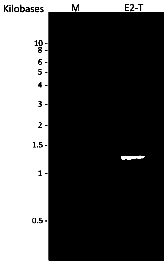
[0064] This example provides the preparation method, purification method and detection of the expressed target protein of classical swine fever virus envelope protein E2 and E0 tetrasubunit oligomeric proteosome.

[0065] 1. Preparation of cell supernatant containing classical swine fever virus envelope protein E2 / E0 tetrasubunit oligomeric protein body

[0066] In this example, the expression and preparation of the four-subunit oligomeric protein body of the envelope protein E2 of classical swine fever virus is illustrated as an example: Insect cells sf-9 are plated in a 6-well cell culture plate, and the recombinant baculovirus The expression plasmid E2-T was transfected, and the cell supernatant was collected six days later to obtain the first-generation recombinant baculovirus seed, which is called the P1 generation. Infect insect cells with the P1 virus seed to amplify the virus seed to obtain the second-generation recombinant baculovirus seed, which is called the P2 gene...

Embodiment 3

[0074] This example provides the detection of aggregation of E2 or E0 tetrasubunit oligomeric proteosomes.

[0075] Envelope proteins E2 and E0 mostly exist in the form of dimers in nature. For the convenience of comparison, an expression plasmid containing E2-6XHis was constructed, prepared and affinity purified according to the above method, and named E2-S. The E2 tetrasubunit oligomeric protein body (tentatively named E2-T here for comparison) and the E2-S protein were denatured and treated under reducing conditions, and Western blots were performed on the samples before and after treatment. Figure 9 The difference between the four-subunit oligomeric proteosome E2-T and the envelope protein E2 dimer in the non-reduced state is clearly shown. It shows that the prepared and purified sample is the E2 tetrasubunit oligomeric proteosome.

[0076] Similarly, the expression plasmid of E0-6XHis was constructed, prepared and affinity purified to obtain E0-6XHis protein, which was...


PUM


Abstract

The invention provides hog cholera virus envelope protein oligomeric protein and a preparation method and application thereof. The oligomeric protein comprises hog cholera virus envelope protein molecules and exogenous oligomerization structural fragments, wherein the hog cholera virus envelope protein molecules are selected from hog cholera virus envelope protein E2 molecules and hog cholera virus envelope protein E0 molecules, the hog cholera virus envelope protein E2 molecules and the exogenous oligomerization structural fragments form the E2 oligomeric protein, and the hog cholera virus envelope protein E0 molecules and the exogenous oligomerization structural fragments form E0 oligomeric protein. The E2 oligomeric protein and the E0 oligomeric protein which serve as the vaccine antigens do not contain any genetic material RNA of hog cholera virus and are safe in production and use. The natural structure and bioactivity of envelope protein in the E2 oligomeric protein and the E0 oligomeric protein are kept, so that the E2 oligomeric protein and the E0 oligomeric protein which serve as the vaccine antigens are evident in immune effect. The oligomeric protein polymerizes the multiple subunit protein of the antigens, overcomes the shortcomings of subunit protein vaccines and is capable of evidently increasing vaccine immunogenicity, high in antigen content and easy in production and purification.

Description

technical field [0001] The present invention relates to the technical fields of bioengineering and virus vaccines, in particular to a preparation method thereof, and a novel vaccine comprising envelope protein E2 and E0 oligomers of classical swine fever virus. As well as the application in preparing preparations for detection of swine fever virus antibodies or preparations for monitoring swine fever virus epidemics. Background technique [0002] Classic Swine Fever (CSF) is an acute, febrile and highly contagious infectious disease of pigs caused by Classic Swine Fever Virus (CSFV), which can be divided into acute, subacute, chronic and Atypical swine fever. Acute swine fever is caused by virulent strains and generally results in high morbidity and mortality in pigs. In addition to causing sepsis, classical swine fever virus can also cause a series of clinical manifestations, such as abortion in pregnant sows, fetal malformation, chronic nutrient consumption, lymphocyte a...

Claims


Application Information

Patent Timeline
Patent Type & Authority Applications(China)
IPC IPC(8): C07K14/18A61K39/12A61P31/14G01N33/569
CPCC07K14/005A61K39/12A61P31/14G01N33/56983C12N2770/24322C12N2770/24334Y02A50/30
Inventor 许雁诺曼·吉利卡李改夏燕
Owner 许雁
Learn More