Preparation method of anti-RET mutant protein monoclonal antibody variable region sequence

A technology of monoclonal antibody and variable region, applied in anti-receptor/cell surface antigen/cell surface determinant immunoglobulin, anti-animal/human immunoglobulin, immunoglobulin, etc. As a result, there were no RET protein point mutations, monoclonal antibodies could not detect protein mutant types, etc., and achieved the effect of convenient and fast screening

Inactive Publication Date: 2020-03-31
安徽环球基因科技有限公司
View PDF0 Cites 0 Cited by
  • Summary
  • Abstract
  • Description
  • Claims
  • Application Information

AI Technical Summary

Problems solved by technology

[0003] Medullary thyroid carcinoma (MTC) can be divided into two types: sporadic medullary thyroid carcinoma (SMTC) and hereditary medullary thyroid carcinoma (HMTC). A large number of studies have confirmed that mutations in the proto-oncogene (RET) are the cause of medullary thyroid carcinoma. The main molecular etiological basis, about 95% of SMTC and more than 80% of HMTC are caused by RET gene mutations, current clinical research is limited to the molecular level based on gene sequencing, and theoretically RET gene mutations will eventually lead to RET protein Amino acid mutations can cause diseases, but there is no specific research on RET protein point mutations;
[0004] Antibody refers to the protective protein produced by the body due to the stimulation of antigens. Antibodies prepared against RET mutant proteins can well inhibit the effect of RET mutant proteins. The traditional way of preparing antibodies is through artificially prepared hybridomas. Cells produce monoclonal antibodies to prepare antibodies. The commonly used monoclonal antibody preparation methods use proteins as antigens to immunize monoclonal antibodies. The prepared monoclonal antibodies cannot detect protein mutants. In monoclonal antibodies During the preparation process, the hybridoma cells are cultured in the form of liquid medium, which is complicated and requires subcloning to produce monoclonal antibodies. ELISA is often used in the screening of hybridoma-positive cell lines, regardless of the ELISA method. There are many interference factors, whether by instrument or manual operation. At the same time, due to the interference of autoantibodies and heterophilic antibodies, false positives are prone to occur and the test results are easily affected, making the production efficiency of monoclonal antibodies low.

Method used


Image

  • Preparation method of anti-RET mutant protein monoclonal antibody variable region sequence
  • Preparation method of anti-RET mutant protein monoclonal antibody variable region sequence
  • Preparation method of anti-RET mutant protein monoclonal antibody variable region sequence

Examples


Embodiment 1

[0035] The preparation method of the variable region sequence of the anti-RET mutant protein monoclonal antibody, the specific steps are as follows:

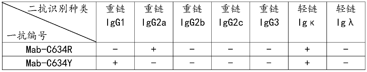
[0036] Step S1: For the C634 site of the RET protein, design and synthesize the original polypeptide C634, mutate the C634 site, and synthesize the mutant polypeptides C634R, C634Y, C634S, C634G, C634W, C634F, and use glutaraldehyde as a cross-linker for the mutant polypeptides respectively. The agent is coupled with the carrier protein KLH to prepare the antigen, and the antigen is mixed to prepare the antigen mixture and emulsified with Freund's adjuvant to obtain the emulsified antigen;

[0037] Step S2: Construct mutant polypeptide expression sequences C634R, C634Y, C634S, C634G, C634W, C634F respectively to the amino terminus of purple fluorescent protein mcherry, and clone them into pet22b expression vector. After prokaryotic expression and purification, mutant proteins C634R-mcherry, C634Y- After mcherry, C634S-mcherry, C...

Embodiment 2

[0042] Antibody WB detection verification, the specific steps are as follows:

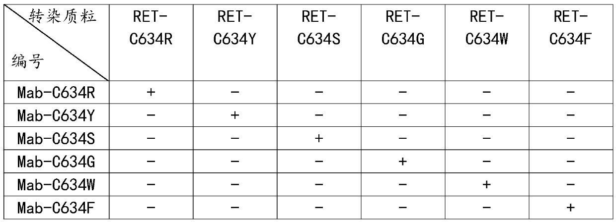
[0043] Step S1: respectively construct pcDNA3.1-RET-C634R, pcDNA3.1-RET-C634Y, pcDNA3.1-RET-C634S, pcDNA3.1-RET-C634G, pcDNA3.1-RET-C634W, pcDNA3.1-RET -C634F full-length protein eukaryotic expression vector, using endotoxin-removing plasmid extraction kit to extract plasmids to obtain 6 kinds of plasmids;

[0044] Step S2: revive HeLa cells, culture HeLa cells in an incubator to log phase, press 1×10 5 Cells / mL were passaged into a 6-well plate, and the addition amount was 2 mL / well. After the 6-well plate was incubated in an incubator for 24 hours, the 6 plasmids prepared in step S1 were respectively transfected with liposome transfection reagent. After reaching Hela cells and culturing for 48 hours, the cells were digested with trypsin, and the cells were collected into EP tubes, washed three times with PBS, and 100 μL of cell lysate was added to the EP tubes. Under the conditions of 4 °C and ...

Embodiment 3

[0051] Antibody IF detection and verification, the specific steps are as follows:

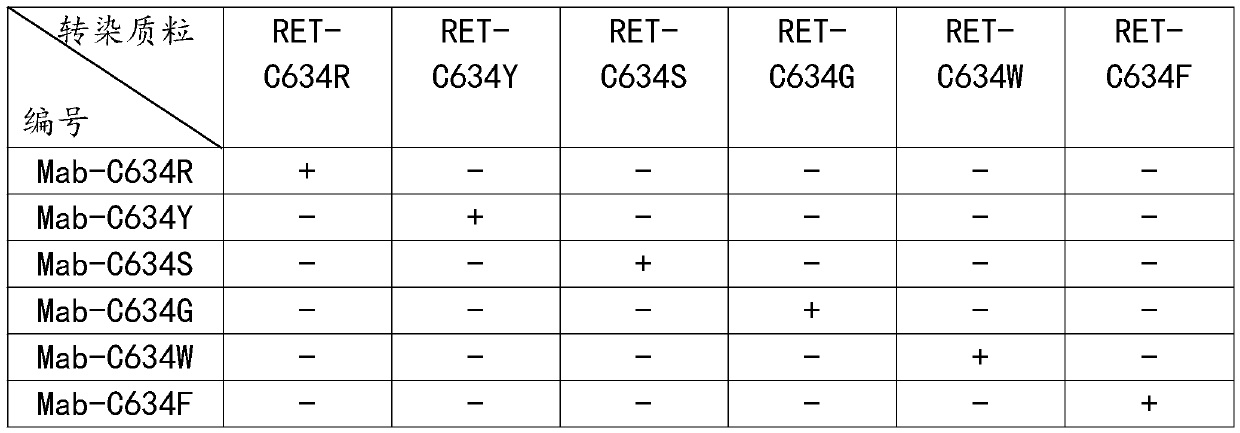
[0052] Step S1: respectively construct pcDNA3.1-RET-C634R, pcDNA3.1-RET-C634Y, pcDNA3.1-RET-C634S, pcDNA3.1-RET-C634G, pcDNA3.1-RET-C634W, pcDNA3.1-RET -C634F eukaryotic expression vector, using endotoxin-removing plasmid extraction kit to extract plasmids to obtain 6 kinds of plasmids;

[0053] Step S2: revive HeLa cells, culture HeLa cells in an incubator to log phase, press 2 × 10 4 Cells / mL were passaged into a 24-well plate, and the addition amount was 1 mL / well. After the 24-well plate was cultured in an incubator for 24 hours, the 6 plasmids prepared in step S1 were respectively transfected with liposome transfection reagent. Transfected into Hela cells and cultured for 48h to obtain transfected cells;

[0054] Step S3: Aspirate the medium in the wells of the 24-plate in step S2, rinse the transfected cells prepared in step S2 twice with PBS, aspirate the PBS, and add 2-PBS prepared to...


PUM


Abstract

The present invention discloses a preparation method of an anti-RET mutant protein monoclonal antibody variable region sequence. Mutation is performed at the C634 position of a RET protein to synthesize a polypeptide sequence, and the polypeptide and a carrier protein KLH are respectively coupled and combined with freund's adjuvant for emulsification to prepare an emulsified antigen, mutants are separately used to construct expression sequences to a purple fluorescent protein mcherry amino terminus to be combined with an expression vector to prepare a detection agent, mice are immunized with the emulsified antigen and mouse myeloma SP2 / 0 cells and spleen cells of the immunized mice are subjected to cell fusion, and are cultured with a semi-solid culture medium and the detection agent, by observing formation of clone balls and color changes of the culture medium, positive clones are selected into new 6-well plates and subjected to expanded culture by a changed liquid culture medium, each cell line is then transferred to a shake flask for culture, a supernatant of the culture medium is collected, antibodies are purified, and the antibodies are sequenced to obtain the variable regionsequence.

Description

technical field [0001] The invention belongs to the field of monoclonal antibody preparation, and in particular relates to a preparation method of the variable region sequence of an anti-RET mutant protein monoclonal antibody. Background technique [0002] The RET proto-oncogene encodes a group of transmembrane proteins belonging to the tyrosine kinase receptor superfamily, namely RET proteins. The receptor tyrosinase plays a role in the connection between the extracellular environment and the nucleus, and the signal is transmitted from the cell surface to the cytoplasm. , and then to the nucleus, the extracellular part of the receptor forms an inter-receptor aggregation after connecting with a special ligand, which promotes the activation of the kinase. The RET gene mutation enhances the function of RET tyrosine kinase signal transduction in many ways. It promotes the activation of kinases and the transformation of proto-oncogenes, and its mutations are closely related to t...

Claims


Application Information

Patent Timeline
Patent Type & Authority Applications(China)
IPC IPC(8): C07K16/28
CPCC07K16/2863C07K2317/56
Inventor 雍金贵缪连军张磊
Owner 安徽环球基因科技有限公司
Why Eureka
Try Eureka