Flow identification nano vesicle used for cell capture, and preparation method and applications thereof

A nanovesicle, cell technology, applied in cell dissociation methods, biochemical equipment and methods, bioreactors/fermenters for specific purposes, etc., can solve problems such as few multivalent effects, and reduce non-specific adsorption , the effect of good monodispersity and low polydispersity index

Inactive Publication Date: 2020-06-05
RENJI HOSPITAL AFFILIATED TO SHANGHAI JIAO TONG UNIV SCHOOL OF MEDICINE
View PDF6 Cites 5 Cited by
  • Summary
  • Abstract
  • Description
  • Claims
  • Application Information

AI Technical Summary

Problems solved by technology

However, the multivalent effects of enhanced fluidity at cell membrane interfaces ...

Method used


Image

  • Flow identification nano vesicle used for cell capture, and preparation method and applications thereof
  • Flow identification nano vesicle used for cell capture, and preparation method and applications thereof
  • Flow identification nano vesicle used for cell capture, and preparation method and applications thereof

Examples


preparation Embodiment 1

[0047] Preparation Example 1 Preparation of flow recognition nanovesicles for cell capture

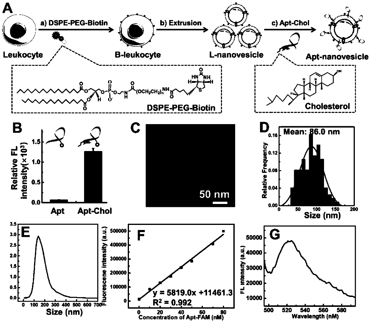
[0048] Such as figure 1 As shown in A, the leukocyte membrane-derived flow-recognition nanovesicles for cell capture were prepared by a top-down method: 1) Mouse macrophage J774A.1 was selected as a model leukocyte, and J774A. 1 Cells were routinely cultured with medium containing DSPE-PEG-biotin (60 μM) for 3 h (attached figure 1 A(a)); 2) Collect the above-mentioned biotinylated J774A.1 cells, use a homogenizer to break the cells to form a homogenate, and centrifuge through a sucrose density gradient (the mass concentration gradient is 30%, 40%, 50% , 28000rpm, 2h) to separate and purify the cell membrane fragments; 3) collect the cell membrane fragments, and use a nano-extruder to extrude the cell membrane fragments into surface biotinized nanovesicles with a particle size of about 100nm (attached figure 1 A(b)); 4) In order to endow biomimetic nanovesicles with tumor cell recogni...

preparation Embodiment 2

[0049] Preparation Example 2 Preparation of Flow Recognition Nanovesicle Modified Microfluidic Chip for Cell Capture

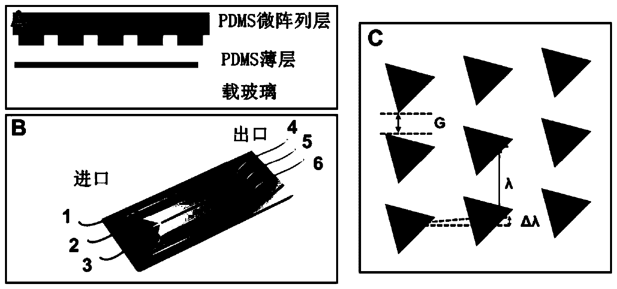
[0050] See attached figure 2 A-C, to make a microfluidic chip, from top to bottom, the PDMS microarray layer, PDMS thin layer, and glass carrier are sequentially bonded with oxygen plasma to form a complete chip. There are three sample inlets (1), (2), (3) on both sides of the chip (1 and 3 are for buffer solution injection, 2 is for whole blood sample injection) and three sample outlets (4), (5) , (6), between the sample inlet and the sample outlet is a triangular microarray, wherein the microarray arrangement adopts the DLD design principle, and the chip arranged by the microcolumn is called DLD-Chip for short, as shown in the attached figure 1 Shown in B.

[0051] In this embodiment, the chip size is designed to be 1 cm wide and 4.5 cm long.

[0052] In this embodiment, the vertical distance G between the pillars is set to 32 μm, the distance λ between ...

experiment Embodiment 1

[0054] Experimental example 1 Selective recognition of flow-recognized nanovesicles for cell capture and EpCAM-positive SW480 tumor cells

[0055] In order to investigate the ability and specificity of the above-mentioned flow recognition nanovesicles for cell capture to recognize tumor cells, EpCAM-positive SW480 cells and control blood cells were treated with 0.1 nM terminal fluorescein-modified nucleic acid aptamer functionalized nanovesicles (Apt-nanovesicles ), 200nM terminal fluorescein-modified nucleic acid aptamer (Apt) was incubated at 4°C for 0.5h. After washing with buffer solution for 3 times, the fluorescence intensity of cells labeled with nanovesicles or aptamers was detected by flow cytometry (at least 10000 cells were counted). At the same time, the above-mentioned cells and fluorescein-modified DNA random sequences were treated according to the above-mentioned steps as a negative control.

[0056] The results show that the nanovesicles prepared in Example 1 ...


PUM

PropertyMeasurementUnit
Dissociation constantaaaaaaaaaa

Abstract

The invention discloses a flow identification nano vesicle used for cell capture. The nano vesicle is characterized in that the nano vesicle includes a sphere structure formed by natural cell membranes or biomimetic membranes through wrapping; and the interfaces of the membranes are modified with specific recognition molecules through physical, chemical or biological action. A method by using thenano vesicle to capture circulating tumor cells is also provided. A microfluidic chip is provided and modified with the nano vesicle; and a method by using the microfluidic chip to capture the circulating tumor cells is provided as well. Through the using of liquidity-enhanced multivalent strategies, the efficient, selective and mild capturing of the circulating tumor cells can be realized.

Description

technical field [0001] The invention relates to the field of interface engineering, in particular to capturing ring tumor cells by using natural biofilm or bionic film. Background technique [0002] Cancer seriously threatens human life and health. According to the latest forecast data released by the World Health Organization, the number of cancer deaths worldwide in 2018 was as high as 9.6 million. The development of precision diagnosis and treatment technology is an important strategy to improve the survival rate of cancer patients. However, tissue biopsy, as the gold standard for tumor diagnosis, has shortcomings such as difficult sampling, strong invasiveness, large deviation, and hysteresis. Therefore, the development of new technologies for precise cancer diagnosis complementary to tissue biopsy is an urgent scientific problem to be solved. [0003] Circulating tumor cells (Circulating Tumor Cells, CTCs) refer to tumor cells that shed from solid tumor tissues and e...

Claims


Application Information

Patent Timeline
IPC IPC(8): C12N5/078C12N5/09B01J13/04C12M1/00B01L3/00
CPCB01J13/046B01L3/5027C12M47/04C12N5/0634C12N5/0641C12N5/0644C12N5/0693C12N2509/00
Inventor 杨朝勇吴玲玲宋彦龄
Owner RENJI HOSPITAL AFFILIATED TO SHANGHAI JIAO TONG UNIV SCHOOL OF MEDICINE
Why Eureka
Try Eureka