Annexin V fluorescent labeling method for detecting early apoptosis of cells

A technology of fluorescent labeling and labeling methods, applied in chemical instruments and methods, biochemical equipment and methods, and fusion with spectrum/fluorescence detection, etc., can solve the problems of size affecting AnnexinV activity, application limitations, and poor photostability of fluorescent proteins

Pending Publication Date: 2020-06-26
DALIAN INST OF CHEM PHYSICS CHINESE ACAD OF SCI
View PDF5 Cites 2 Cited by
  • Summary
  • Abstract
  • Description
  • Claims
  • Application Information

AI Technical Summary

Problems solved by technology

Among them, the organic fluorescent dye is labeled by coupling reaction with the side chain amino group on the Annexin V protein, but this reaction is not selective, and it is difficult to control the labeled site on Annexin V and the number of fluorescent dye molecules connected.
Labeling with more than two fluorochrome molecules leads to inactivation of Annexin V, study shows
It is difficult to implement a fluorescent single-molecule labeling process that does not affect the...

Method used


Image

  • Annexin V fluorescent labeling method for detecting early apoptosis of cells
  • Annexin V fluorescent labeling method for detecting early apoptosis of cells
  • Annexin V fluorescent labeling method for detecting early apoptosis of cells

Examples


Embodiment 1

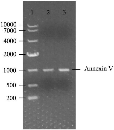
[0036] (1) Acquisition of Annexin V target gene:

[0037] Primer design: Design the upstream and downstream primers of the Annexin V target fragment and introduce the Nhe1 restriction site (GCTAGC), and introduce the protective bases TACTA and ACTA in the upstream of the restriction site. The primers are as follows:

[0038] Upstream primer 5’TACTAGCTAGCATGGCACAGG TTCTCAGAGG 3’

[0039] Downstream primer 5’ACTAGCTAGCGTCATCTTCT CCACAGAGCA G 3’

[0040] PCR amplification of Annexin V target gene: PCR reaction system 50μL: PCR buffer (10×) 5μL; dNTP (10×) 5μL; pCMV-Annexin V 1μL; upstream and downstream primers (20nM) each 1μL; pfu DNA polymerase 0.5μL; 36.5μL of sterilized double distilled water. The PCR reaction conditions were as follows: 95°C 30sec; [95°C 30sec; 56°C 1min; 68°C 4min;] 25 cycles; 68°C 3min; 4°C incubation. After PCR, run 0.8% agarose nucleic acid electrophoresis, use PCR purification kit to purify the target fragment, use NheI restriction enzyme digestion to expose ...

Embodiment 5

[0054] Fluorescence imaging experiment of the SNAP-DAC fluorescently labeled SNAP-Annexin V fusion protein prepared in Example 5 on early apoptotic cells

[0055] SNAP-DAC fluorescently labeled SNAP-Annexin V fusion protein was prepared into a 2μM mother solution for later use.

[0056] Spread Hela cells (proliferating epidermal cancer cells) in a petri dish. The dish contains 1 mL of DMED high glucose medium containing 10% fetal bovine serum, and culture it at 37°C and 5% carbon dioxide until the cell density is about 40%. Replace Fresh culture medium containing 50μg / mL cell apoptosis inducer NCTD was cultured for 12 hours. Then wash 2 times with PBS and add 2mM CaCl 2 And 0.1μM SNAP-DAC fluorescently labeled SNAP-Annexin V fusion protein, detected under 488nm excitation Figure 5 .

[0057] The fluorescence imaging image of SNAP-Annexin V fusion protein on early apoptotic cells is as follows Figure 5 Shown: Figure 5 -a is bright-field Hela cell imaging, Figure 5 -b is fluoresce...

Embodiment 3

[0059] Fluorescence imaging experiment of SNAP-Cell 505-Star fluorescently labeled SNAP-Annexin V fusion protein prepared in Example 5 on early apoptotic cells

[0060] Place Hela cells (proliferating epidermal cancer cells) in a petri dish. The dish contains 1 mL of DMED high-glycemic medium containing 10% fetal bovine serum, and culture it at 37°C and 5% carbon dioxide until the cell density is about 50%. Replace Fresh culture medium containing 50μg / mL cell apoptosis inducer NCTD was cultured for 12 hours. Then wash 2 times with PBS and add 2mM CaCl 2 And 0.1μM SNAP-Cell 505-Star fluorescently labeled SNAP-Annexin V fusion protein, and perform fluorescence imaging under 488nm excitation. Get with Figure 5 Similar HeLa cell imaging proved the effect of SNAP-Cell 505-Star fluorescently labeled SNAP-Annexin V fusion protein on early apoptosis cells.


PUM

PropertyMeasurementUnit
Molecular weightaaaaaaaaaa

Abstract

The invention provides an Annexin V fluorescent labeling method for detecting early apoptosis of cells. The specific labeling method comprises the following steps: (1) constructing a pET-22b-SNAP-Annexin V gene fusion plasmid vector; (2) performing prokaryotic expression and purifying pET-22b-SNAP-Annexin V fusion protein; and (3) labeling the fusion protein by using fluorescent small molecules and performing purification. Annexin V protein is one of best phosphatidylserine (PS) binding receptors found at present, shows high selectivity and high binding ability for PS binding, and fluorescently labeled AnnexinV is one of most commonly used reagents for detecting the early apoptosis of the cells. The invention creates the novel method of Annexin V fluorescent labeling, firstly, SNAP tag protein and the Annexin V protein are fused, expressed and purified through the genetic engineering method, then a small molecule fluorescent probe connected to a BG group of a special substrate of the SNAP protein is used to label the fusion protein and label the Annexin V, and the apoptotic cells are detected by cell imaging or flow cytometry. The method has important significance for the researchof the cell apoptosis process.

Description

Technical field [0001] The invention belongs to the field of biological analysis and detection, and specifically relates to a method for detecting the AnnexinV fluorescent labeling of early cell apoptosis. Background technique [0002] Apoptosis is the programmed death of organisms to maintain the stability of the internal environment. Abnormal cell apoptosis can lead to many diseases such as tumors, neurodegeneration and autoimmunity. Based on the characteristics of in-situ, real-time and high sensitivity, fluorescence detection of apoptosis can trace the process of apoptosis, help analyze the mechanism of abnormal apoptosis-related diseases, screen new drugs and establish effective treatment methods. In normal cells, phosphatidylserine (PS) is located on the inner side of the cytoplasmic membrane. In the early stage of apoptosis, PS flips from the inner side of the cytoplasmic membrane to the outer side of the cytoplasmic membrane, which is an important signal for the onset of...

Claims


Application Information

Patent Timeline
IPC IPC(8): C12N15/62C12N15/70C07K19/00C12Q1/02
CPCC07K14/47C12N15/70G01N33/5005C07K2319/60
Inventor 徐兆超苗露乔庆龙
Owner DALIAN INST OF CHEM PHYSICS CHINESE ACAD OF SCI
Why Eureka
Try Eureka