Probe for capturing hirudin polypeptide and application of probe

A technology of hirudin and probe, which is applied in the preparation method of peptides, from leech inhibitors, peptides, etc., can solve the problems of low quantitative sensitivity, poor quantitative repeatability, and difficult detection by time method, and achieve good application prospects and high efficiency. high effect

Active Publication Date: 2020-07-24
CHENGDU UNIV OF TRADITIONAL CHINESE MEDICINE
View PDF14 Cites 3 Cited by
  • Summary
  • Abstract
  • Description
  • Claims
  • Application Information

AI Technical Summary

Problems solved by technology

[0003] At present, the content determination of hirudin peptides basically adopts biological activity methods, mainly including thrombin titration, chromogenic substrate method, light scattering method, fibrinogen plate method, thrombin time method, from the pharmaceutical quality control mode From the perspective of applicability, biological detection technology is often more practical than chemical composition detection because it is closely related to safety and effectiveness, but it also has its own defects: a) The quantitative sensitivity is low. When the concentration of the target polypeptide is very low, the titration b) Quantitative repeatability is poor, and the measurement results are easily affected by factors such as free thrombin activity, ambient temperature and substrate concentration
It has been reported that thrombin is coupled to magnetic particles with an amino terminal, and hirudin is isolated and purified from the leaching solution of dried leeches (Application of thrombin-coupled magnetic particle technology to the detection of antithrombin activity in leech medicinal materials, Chinese Medicine Biotechnology Application Journal, 2002), but this method has obvious disadvantages: that is, the connection between thrombin and the amino terminal is connected by a covalent bond (generally -CO-NH-), and the site where the former binds to hirudin may be blocked Blocked or semi-blocked at the amino-terminus of magnetic particles, so the efficiency of binding hirudin is low

Method used


Image

  • Probe for capturing hirudin polypeptide and application of probe
  • Probe for capturing hirudin polypeptide and application of probe
  • Probe for capturing hirudin polypeptide and application of probe

Examples


Embodiment 1

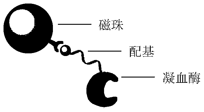
[0040] Preparation of Example 1 probe (3 μm polystyrene carboxyl-ligand-bovine thrombin)

[0041] Step 1: Conjugate streptavidin. 50mg polystyrene carboxyl magnetic beads (3μm, figure 1 ), 5mg EDC and 1mg streptavidin were dispersed in 1mL MES buffer solution, rotated and mixed at 30°C, reacted for 6 hours, separated the reaction product with a magnet, redispersed into 0.1% BSA solution, and sealed for 6 hours , and washed 5 times with PBS buffer to prepare 3 μm streptavidin-modified polystyrene magnetic beads.

[0042] The polystyrene magnetic beads refer to the cores of porous polystyrene microspheres, the magnetic particles are embedded in the pores, the outer layer is coated with polyacrylic acid, and carboxyl groups are distributed on the surface.

[0043] Step 2: Modify the thrombin ligand. The magnetic beads prepared in step 1 were mixed with the ligand at a ratio of 50 mg: 1 μmol, dispersed in PBS buffer, shaken at room temperature for 1 hour, and the product was w...

Embodiment 2

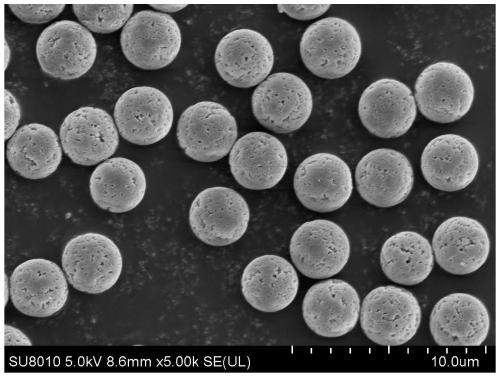
[0046] Preparation of Example 2 Probe (10 μm agarose carboxyl-ligand-bovine thrombin)

[0047] Step 1: Conjugate streptavidin. 100mg agarose carboxyl magnetic beads (10μm, figure 2 ), 5mg EDC and 2mg streptavidin were dispersed into 1mL MES buffer solution, rotated and mixed at 30°C, reacted for 6 hours, separated the reaction product with a magnet, redispersed into 0.1% BSA solution, and blocked for 6 hours Finally, wash with PBS buffer 5 times to prepare 10 μm streptavidin-modified agarose magnetic beads.

[0048] The agarose carboxyl magnetic beads refer to magnetic beads with carboxyl groups sealed by agarose.

[0049] Step 2: Modify the thrombin ligand. Mix the magnetic beads prepared in step 1 with the ligand at a ratio of 100 mg: 2 μmol, disperse them in PBS buffer, shake at room temperature for 1 hour, wash the product with deionized water 5 times, and obtain 10 μm ligand-modified agarose magnetic beads .

[0050] The thrombin ligand is as follows: 5'biotin-TTTTTTT...

Embodiment 3

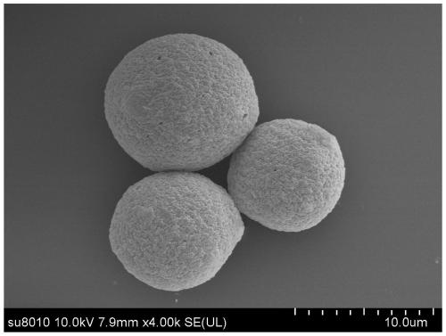
[0052] Preparation of Example 3 Probe (25 μm agarose NHS-ligand-bovine thrombin)

[0053] Step 1: Conjugate streptavidin. 100mg agarose NHS magnetic beads (25μm, image 3 ) and 2 mg of streptavidin were dispersed in 1 mL of MES buffer solution, rotated and mixed at 30°C, and reacted for 6 hours. The reaction product was separated with a magnet, re-dispersed into 0.1% BSA solution, and blocked for 6 hours. Wash with PBS buffer 5 times to prepare 25 μm streptavidin-modified agarose magnetic beads.

[0054] The agarose NHS magnetic beads refer to magnetic beads sealed by agarose with NHS groups (belonging to activated carboxyl groups).

[0055] Step 2: Modify the thrombin ligand. Mix the magnetic beads prepared in step 1 with the ligand at a ratio of 100 mg: 2 μmol, disperse in PBS buffer, shake at room temperature for 1 hour, and wash the product with deionized water 5 times to obtain 25 μm ligand-modified agarose magnetic beads .

[0056] The thrombin ligand is as follows:...


PUM


Abstract

The invention discloses a preparation method and application of a biological recognition probe for capturing hirudin polypeptide. Ligands are modified on the surface of a magnetic solid-phase carrier,thrombin is indirectly fixed, the biological activity is reserved to a great extent, and the obtained probe can be combined with multiple detectors to achieve extraction and determination of the hirudin polypeptide, and has the advantages of being high in selectivity, rapid, sensitive, good in stability, wide in applicability, small in interference, high in repeated utilization rate, environmentally friendly and the like.

Description

technical field [0001] The invention belongs to the field of polypeptide detection. Background technique [0002] Hirudin peptides are bivalent direct thrombin inhibitors with the same or similar chemical structure as natural hirudin and a molecular weight of 5-7 kDa, which can simultaneously act on the active site and substrate recognition site of thrombin. Hirudin is a single-chain polypeptide compound composed of 65-66 amino acids isolated and purified from leech saliva, including three variants of HV 1, HV 2, and HV 3. It is the most effective natural specific inhibitor of thrombin so far. Agents, similarly there are hirudin extracted from Hirudo phenantha and hirudin isolated from leech. The production of natural hirudin is less, which is not conducive to clinical promotion. Recombinant hirudin produced by genetic engineering technology, such as lepirudin and desirudin, have an ideal clinical effect. Studies have shown that hirudin peptides have stronger anticoagulant...

Claims


Application Information

Patent Timeline
IPC IPC(8): C07K14/815C07K1/14G01N33/68
CPCC07K14/815G01N33/68G01N2333/815
Inventor 曹秀君国锦琳聂应罗莎杰张佳斌
Owner CHENGDU UNIV OF TRADITIONAL CHINESE MEDICINE
Why Eureka
Try Eureka