Nanoscale single exosome separation method

A separation method and nanoscale technology, applied in cell dissociation methods, biochemical equipment and methods, treatment of microorganisms with electricity/wave energy, etc., can solve problems such as low separation efficiency, reduced quality, and low dispersion efficiency, and achieve separation The effect of high yield, convenient method and low cost

Active Publication Date: 2020-08-11
XI AN JIAOTONG UNIV
View PDF11 Cites 3 Cited by
  • Summary
  • Abstract
  • Description
  • Claims
  • Application Information

AI Technical Summary

Problems solved by technology

Ultracentrifugation is the most commonly used exosome purification method. Vesicle particles of different sizes are precipitated and purified from soluble molecules by high-speed centrifugation, but repeated centrifugation is likely to cause damage to exosome vesicles and reduce their quality.
The ultrafiltration centrifugation method uses the difference in relative molecular mass to selectively separate samples. This method is simple and efficient, and does not affect the biological activity of exosomes. However, the filter pores are easily blocked, resulting in shortened membrane life and low separation efficiency.
Based on the principles of viscoelastic flow and nanowire traps, the microchannel method realizes the effective separation of exosomes of different scales through acoustic, electrical and electromagnetic manipulation. This method has the advantages of minimal consumption, high resolution and low cost. , but the diameter of the microchannel is usually above the micron level, making it difficult to separate single exosomes at the nanoscale
[0004] Magnetic bead immunoassay is an immunoaffinity technique. The protein surface of exosomes has specific receptors, and different types of exosomes are captured from the sample by nanoparticles with corresponding modified antibodies, so as to achieve exosome Selective separation, this method has the advantages of high specificity and high-purity exosomes, but lacks appropriate carrier support to separate independent single exosomes, resulting in the efficie

Method used


Image

  • Nanoscale single exosome separation method
  • Nanoscale single exosome separation method
  • Nanoscale single exosome separation method

Examples


Embodiment 1

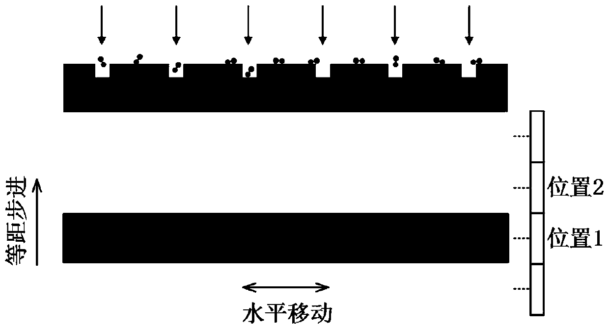
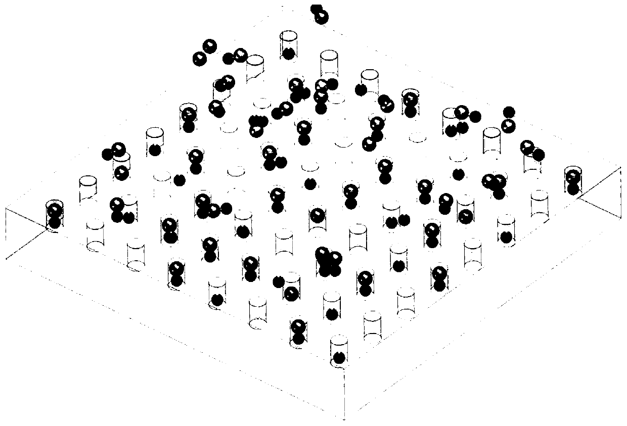
[0066] In this embodiment, the diameter of the nanoholes in the nanohole microarray structure is set to 100±15 nm, and the depth of the nanoholes is set to 250±30 nm. The horizontal and vertical centers of the nano-holes are equally spaced at 2 μm to form a 201×201 lattice square microarray. The number of nano-holes on a single sheet is 40,401, and they are located in the central area of ​​the general-purpose cover glass. The nanohole microarray structure is prepared using the femtosecond laser direct writing equipment of the cooperative research group.

[0067] Dilute the exosome-containing cell culture medium after biological extraction and purification to a concentration of 1×10 NTA quantitative test 7 pieces / ml.

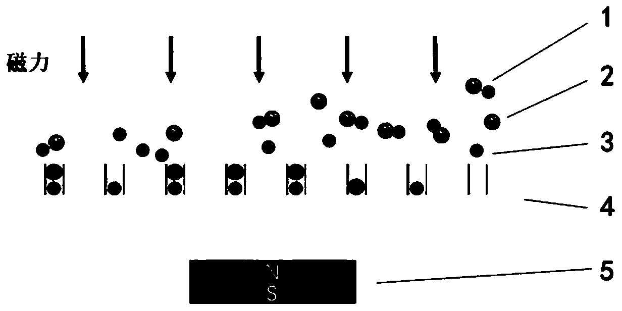
[0068] Choose common and consistent superparamagnetic amino magnetic beads with a diameter of 50nm. The surface contains amino functional groups that can specifically couple with exosome coating proteins. Dilute the magnetic bead sample solution to 1×10 7 ~1×10...

Embodiment 2

[0078] In this embodiment, the diameter of the nanoholes in the nanohole microarray structure is set to 200±15 nm, and the depth of the nanoholes is set to 250±30 nm. The horizontal and vertical centers of the nano-holes are equally spaced at 2 μm to form a 201×201 lattice square microarray. The number of nano-holes on a single sheet is 40,401, and they are located in the central area of ​​the general-purpose cover glass. The nanohole microarray structure is prepared using the femtosecond laser direct writing equipment of the cooperative research group.

[0079] Dilute the exosome-containing cell culture medium after biological extraction and purification to a concentration of 1×10 NTA quantitative test 8 pieces / ml.

[0080] Use common, consistent, superparamagnetic amino beads with a diameter of 100nm, the surface of which contains amino functional groups that can specifically couple with exosome coating proteins, and dilute the magnetic bead sample solution 1×10 8 ~1×10 9...

Embodiment 3

[0090] In this embodiment, the diameter of the nanoholes in the nanohole microarray structure is set to 250±15 nm, and the depth of the nanoholes is set to 300±30 nm. The horizontal and vertical centers of the nano-holes are equally spaced at 2 μm to form a 201×201 lattice square microarray. The number of nano-holes on a single sheet is 40,401, and they are located in the central area of ​​the general-purpose cover glass. The nanohole microarray structure is prepared using the femtosecond laser direct writing equipment of the cooperative research group.

[0091] Dilute the exosome-containing cell culture medium after biological extraction and purification to a concentration of 1×10 NTA quantitative test 9 pieces / ml.

[0092] Choose common and consistent superparamagnetic amino magnetic beads with a diameter of 120nm, the surface of which contains amino functional groups that can specifically couple with exosome coating proteins, and dilute the magnetic bead sample solution to...


PUM


Abstract

The invention discloses a nanoscale single exosome separation method. The method comprises the following steps of fully diluting a cell culture solution containing exosome; adding a superparamagneticamino magnetic bead diluent, and carrying out low-frequency oscillation mixing until the nano magnetic beads are coupled with the exosome; transferring a coupling solution to a surface layer of a nanopore array microchip; placing the microchip on a shaking table, and oscillating in low speed and small angle, so that the coupling solution is uniformly distributed on the surface layer of the chip; placing a magnetic source on the lower surface of the microchip, and enabling a magnetic bead exosome couplet to enter a nanopore array by using a progressive magnetic adsorption mode; under the actionof larger magnetic force, maintaining stable adsorption of the magnetic bead exosome couplet, cleaning the upper surface of a substrate with a buffer solution, and standing to obtain the nano microchip containing the single exosome which can be directly used for subsequent detection. On the basis of the nanopore microarray microchip with the high-precision screening effect, high-purity single exosome can be obtained through magnetic bead coupling exosome and progressive magnetic adsorption.

Description

technical field [0001] The invention belongs to the technical field of single exosome analysis, and in particular relates to a nanoscale single exosome separation method. Background technique [0002] Exosomes refer to biologically active vesicles with a diameter of about 30-150 nm secreted by (most) cells. Exosomes have a typical lipid bilayer membrane structure, carrying important information such as various proteins, lipids, and nucleic acids secreted by the mother cell, and are important carriers and key messengers for information exchange between cells. It plays an important role in the physiological and pathological processes of various diseases. [0003] Currently, exosome isolation techniques mainly include ultracentrifugation, ultrafiltration centrifugation, microchannel method, magnetic bead immunoassay, etc. Ultracentrifugation is the most commonly used exosome purification method. Vesicle particles of different sizes are precipitated and purified from soluble m...

Claims


Application Information

Patent Timeline
IPC IPC(8): C12N5/00C12N13/00
CPCC12N5/00C12N13/00C12N2509/00C12N2509/10
Inventor 黄辰刘涛李国卿何韬杨树明童东东
Owner XI AN JIAOTONG UNIV
Why Eureka
Try Eureka