
Mitochondrion-lysosome migration type membrane potential fluorescent probe CSP

A fluorescent probe, membrane potential technology, applied in fluorescence/phosphorescence, luminescent materials, material analysis by optical means, etc., can solve the problems of limited detection sensitivity, automatic acquisition, practical operation and inconvenience of detection, etc., to achieve real-time Tracking, good reversibility effects

Active Publication Date: 2020-10-16
SHANXI UNIV
View PDF4 Cites 1 Cited by
  • Summary
  • Abstract
  • Description
  • Claims
  • Application Information

AI Technical Summary

Problems solved by technology

Among them, the detection sensitivity of the quenching type probe is limited due to the high background signal; the mitochondrial-nucleus migration type probe evaluates the membrane potential by calculating the ratio of the fluorescence intensity of the nucleus to the whole cell, which cannot be directly measured by the laser confocal microscope. The imaging software is automatically obtained, which undoubtedly brings great inconvenience to actual operation and detection

Method used


Image

  • Mitochondrion-lysosome migration type membrane potential fluorescent probe CSP
  • Mitochondrion-lysosome migration type membrane potential fluorescent probe CSP
  • Mitochondrion-lysosome migration type membrane potential fluorescent probe CSP

Examples


Embodiment 1

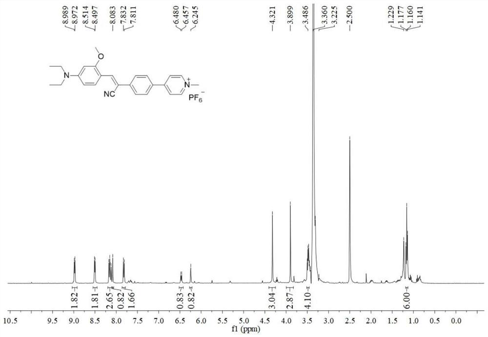
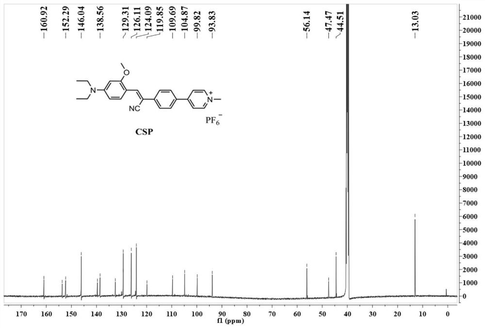
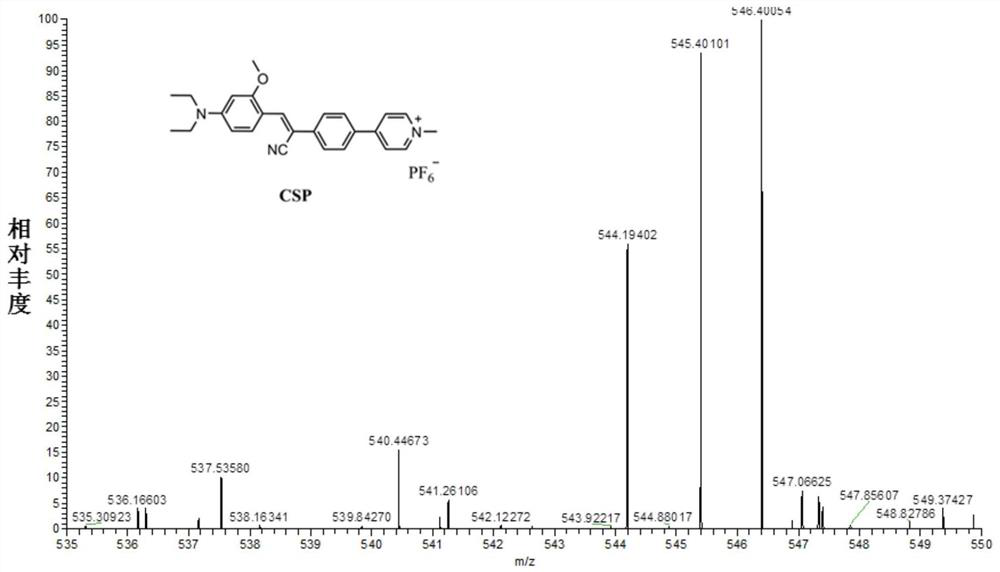
[0032] Preparation and characterization of a mitochondrial-lysosome migratory membrane potential fluorescent probe (CSP) with aggregation-induced emission properties:

[0033]

[0034] (1) In a round bottom flask, 4-bromophenylacetonitrile (0.588g, 3mmol) and t-BuOK (0.336g, 3mmol) were successively added to (30mL) absolute ethanol, and stirred at room temperature for 10 minutes; then 4-Diethylamino-2-methoxy-benzaldehyde (0.621g, 3mmol) was slowly added to the above mixture, and refluxed for 6 hours; the system was cooled to room temperature, the solvent was concentrated in vacuo, and purified by silica gel column chromatography (petroleum ether / ethyl acetate, 5:1, v / v) to obtain compound 2 (0.806 g, 70%) as a yellow solid. 1 H NMR (400MHz, CDCl 3 ):δ(ppm):8.26(d,J=8.8Hz,1H),7.91(s,1H),7.53–7.48(m,4H),6.36(dd,J=9.2,2.0Hz,1H),6.11 (s, 1H), 3.87 (s, 3H), 3.43 (q, J=6.8Hz, 4H), 1.23 (t, J=7.2Hz, 6H).

[0035] (2) Compound 2 (0.346g, 0.9mmol), K2 CO 3 (0.138g, 1mmol) and 4...

Embodiment 2

[0038] The near-infrared aggregation-induced fluorescence emission characteristics of the probe CSP in this example in acetonitrile / water mixed solvent:

[0039] The probe was diluted with acetonitrile / water mixed solvent to a final concentration of 5 μmol / L, the excitation wavelength was fixed at 475 nm, and the fluorescence emission spectrum of the probe changed with water content was recorded ( Figure 4 ), and plot the probe relative fluorescence intensity (I / I 0 ) in the acetonitrile / water mixed system with the curve of water content ( Figure 5 ). As the water volume ratio increased from 0% to 95%, the fluorescence intensity at 678nm increased sequentially, and reached the maximum when the water volume was 85%, indicating that the probe had typical aggregation-induced luminescence characteristics. Analysis of the standard by dynamic light scattering shows that the probe has formed an aggregated state with a hydration diameter of 135.1 nm in acetonitrile / water mixed sol...

Embodiment 3

[0041] The solid-state fluorescence emission performance of the probe CSP of this embodiment

[0042] The excitation wavelength was fixed at 475nm, and the solid-state fluorescence emission spectrum of the probe was recorded. Such as Figure 7 As shown, the maximum fluorescence emission intensity of the solid-state probe is 672nm, which is in the near-infrared emission range (fluorescence emission>650nm), indicating that the probe has aggregation-inducing properties of near-infrared emission. At the same time, the Stoke shift is as high as 197nm, which can effectively reduce the interference of excitation light. In addition, the solid-state probe exhibits bright red fluorescence under UV light (inset); the calculated solid-state fluorescence quantum yield of the probe is 9.8%.


PUM


Abstract

The invention discloses a mitochondrion-lysosome migration type membrane potential fluorescent probe CSP, and belongs to the technical field of mitochondrion membrane potential fluorescent probes. Themitochondrion-lysosome migration type membrane potential fluorescent probe CSP has the near-infrared aggregation-induced fluorescence emission characteristic, and the positive charge structure of themitochondrion-lysosome migration type membrane potential fluorescent probe CSP is combined with the electrostatic interaction of mitochondrion negative membrane potential, so that the probe is aggregated in mitochondria in a targeted manner; the membrane potential is reduced; the probe is released from mitochondria and migrates to lysosome; the probe and the commercially available lysosome specific dye are subjected to co-localization fluorescence imaging, and the characteristic that the co-localization coefficient of the probe and the commercially available lysosome specific dye is increasedalong with reduction of membrane potential is utilized, so that real-time tracking of mitochondrial membrane potential in normal living cells is realized, and visual distinguishing application of dead living cells is realized.

Description

technical field [0001] The invention belongs to the technical field of mitochondrial membrane potential fluorescent probes, in particular to a mitochondrial-lysosome migration type membrane potential fluorescent probe CSP. Background technique [0002] During the mitochondrial tricarboxylic acid cycle, the protons inside the mitochondria will be actively transported to the outside of the mitochondria, thereby forming a mitochondrial membrane potential (-160mV ~ -180mV) that is negative inside and positive outside, which is an important indicator that reflects mitochondrial metabolic function and cell activity. parameter. Abnormal mitochondrial membrane potential can lead to mitochondrial dysfunction, which is often closely related to some cancers, obesity and Parkinson's disease. In addition, numerous studies have shown that the reduction of mitochondrial membrane potential is considered to be one of the earliest events in the process of apoptosis cascade, even before the e...

Claims


Application Information

Patent Timeline
IPC IPC(8): C07D213/57C09K11/06G01N21/64
CPCC07D213/57C09K11/06C09K2211/1029G01N21/6428
Inventor 樊丽王晓东张跃伟李峰董川
Owner SHANXI UNIV
Features
  • Generate Ideas
  • Intellectual Property
  • Life Sciences
  • Materials
  • Tech Scout
  • Unparalleled Data Quality
  • Higher Quality Content
  • 60% Fewer Hallucinations
Learn More