Extracellular vesicle separation and enrichment method based on anionic polymer modified matrix

A separation and enrichment, anion technology, applied in the field of extracellular vesicle separation and enrichment, can solve the problems of small capture capacity, high reagent cost, extracellular vesicle damage, etc., to achieve less free DNA pollution, high purity of the isolated product, Simple effect of separation process

Active Publication Date: 2020-12-08
HANGZHOU ZIJING BIOLOGY CO LTD
View PDF6 Cites 3 Cited by
  • Summary
  • Abstract
  • Description
  • Claims
  • Application Information

AI Technical Summary

Problems solved by technology

Ultracentrifugation is widely used in the separation of extracellular vesicles, but the separation process requires a large ultracentrifuge, and a large amount of impurity proteins and nucleic acids are inevitably co-separated, resulting in low purity of the isolated extracellular vesicles. The difference in vesicle size and density will easily lead to a series of problems such as the discarding of some extracellular vesicles with lower density and smaller particle size, and the inevitable damage of extracellular vesicles caused by excessive centrifugal force 13
Although

Method used


Image

  • Extracellular vesicle separation and enrichment method based on anionic polymer modified matrix
  • Extracellular vesicle separation and enrichment method based on anionic polymer modified matrix
  • Extracellular vesicle separation and enrichment method based on anionic polymer modified matrix

Examples


Embodiment 1

[0043] Example 1. Sodium alginate modified glass fiber is used for the separation of extracellular vesicles in cell culture medium

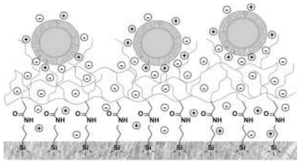
[0044] Sodium alginate modified glass fiber spin column is made by fixing sodium alginate to the surface of glass fiber by chemical modification. The first is the activation of the glass fiber surface, using a mixture of concentrated sulfuric acid (98%) and hydrogen peroxide in a volume ratio of 7:3 as the activation reagent. Put the filter paper into a glass vessel that has been repeatedly cleaned, put the filter paper into the bottom of the vessel, add concentrated sulfuric acid, then add hydrogen peroxide in proportion under shaking conditions, react at room temperature for 30 minutes, fully wash with deionized water, and then dry. The activated glass fiber was added to the APTES solution with a mass fraction of 1%, reacted at room temperature for 30 minutes, fully washed with deionized water three times, and dried at 80°C for more than 2 hour...

Embodiment 2

[0047] Example 2. Heparin-modified glass fiber for the separation of extracellular vesicles in plasma

[0048] The production of heparin-modified glass fiber spin column uses chemical modification to fix heparin on the surface of glass fiber. The first is the activation of the glass fiber surface, using a mixture of concentrated sulfuric acid (98%) and hydrogen peroxide in a volume ratio of 7:3 as the activation reagent. Put the filter paper into a glass vessel that has been repeatedly cleaned, put the filter paper into the bottom of the vessel, add concentrated sulfuric acid, then add hydrogen peroxide in proportion under shaking conditions, react at room temperature for 30 minutes, fully wash with deionized water, and then dry. Add the activated glass fiber into 1% APTES solution, react at room temperature for 30 minutes, wash thoroughly with deionized water three times, and dry at 80°C for more than 2 hours. Mix heparin (10mg / ml) and EDC (0.8mg / ml) at a volume ratio of 1:1...

Embodiment 3

[0051] Example 3. Heparin-modified magnetic beads used for the separation of extracellular vesicles in urine

[0052] The production of heparin-modified magnetic beads uses chemical modification to immobilize heparin on the surface of the magnetic beads. The first is the activation of the surface of the magnetic beads, using a mixture of concentrated sulfuric acid (98%) and hydrogen peroxide in a volume ratio of 7:3 as an activation reagent. Put the magnetic beads into a glass vessel that has been repeatedly cleaned, put the filter paper into the bottom of the vessel, add concentrated sulfuric acid, then add hydrogen peroxide in proportion under shaking conditions, react at room temperature for 30 minutes, fully wash with deionized water, and then dry . The activated magnetic beads were added to 1% APTES solution, reacted at room temperature for 30 minutes, fully washed with deionized water three times, and dried at 80°C for more than 2 hours. Mix heparin (10mg / ml) and EDC (...


PUM


Abstract

The invention discloses a simple, low-cost and high-universality extracellular vesicle separation and enrichment method. The method is characterized in that extracellular vesicles in sample types suchas a culture medium, plasma, serum, urine, saliva, emulsion, pleural effusion and cerebrospinal fluid are separated and enriched through electrostatic interaction. According to the implementation mode of the method, the extracellular vesicles are captured by utilizing an anionic polymer modified matrix material under an acidic condition, and the extracellular vesicles captured in the previous step are eluted under a neutral or alkaline condition, so that the purpose of separating and enriching the extracellular vesicles is finally achieved. Compared with an existing extracellular vesicle separation and enrichment method, the method has the advantages of being simple in separation process, low in cost, good in universality and high in downstream detection compatibility.

Description

technical field [0001] The invention belongs to the field of in vitro diagnosis and targeted drug loading, and in particular relates to a method for separating and enriching extracellular vesicles. Background technique [0002] Extracellular vesicles are a kind of particles secreted by cells into the extracellular space and wrapped by phospholipid bimolecular membranes, with a diameter between 30nm and 2000nm. As an intercellular communication carrier, it is widely distributed in human body fluids such as blood, urine, pleural effusion, saliva, ascites, milk, nasal mucus and cerebrospinal fluid, etc. 2,3 . A large number of studies have proved that extracellular vesicles carry effector molecules such as various proteins, lipids, DNA, mRNA, microRNA, and non-coding RNA, and they communicate with neighboring cells and distant cells through these effector molecules. communication and material exchange 1,4-6 . In recent years, more and more studies have confirmed that extrac...

Claims


Application Information

Patent Timeline
IPC IPC(8): C12N5/00
CPCC12N5/00C12N2509/00
Inventor 刘鹏李尚霖
Owner HANGZHOU ZIJING BIOLOGY CO LTD
Why Eureka
Try Eureka