Preparation and application of a type I hyaluronidase fluorescent nanosensor
A hyaluronidase and nanosensor technology, which is applied in the fields of nanotechnology and analysis and detection to achieve the effects of simple operation, good social value and application prospects, and high specificity
- Summary
- Abstract
- Description
- Claims
- Application Information
AI Technical Summary
Problems solved by technology
Method used
Image

Examples
Embodiment 1
[0031] Example 1: Preparation and characterization of nanosensors
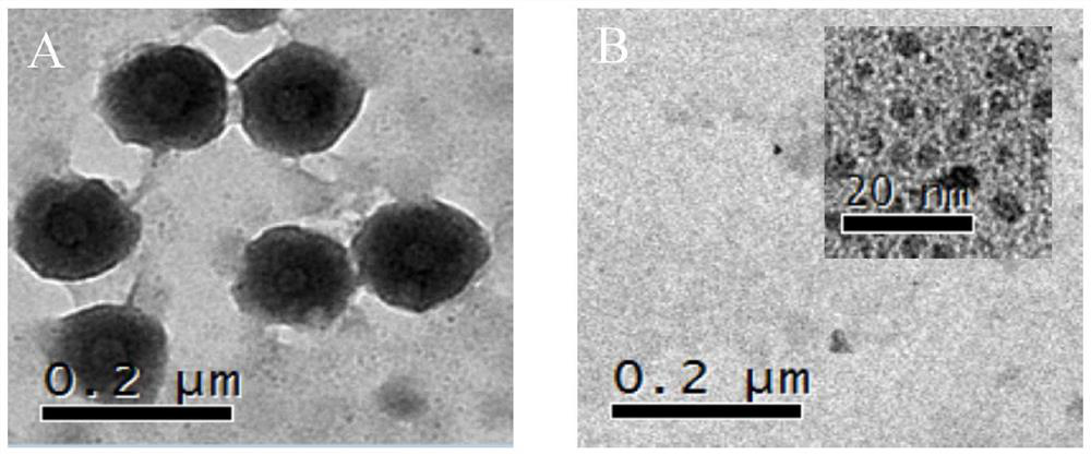
[0032] 1 mg of TPC3 dye and 2 mg of CHA polymer were added to 1 ml of DMSO, and 3 ml of ultrapure water was slowly added dropwise to the solution under magnetic stirring at 37 °C for 30 min, and white micelle particles gradually appeared in the solution. After the reaction, the unwrapped TPC3 dye molecules were removed from the reaction solution with a dialysis bag with a molecular weight cut-off of 3500 KD, and dialyzed for 3 days. During this process, the white micellar particles were slowly dispersed in the dialysis bag until they disappeared to obtain a uniform size TPC3@CHA micellar solution. The final product was stored at 4°C. Depend on figure 1 (A) The TEM image shows that the size of TPC3@CHA is about 160 nm, and there is no agglomeration phenomenon, and the uniformity is good.
Embodiment 2
[0033] Example 2: Titration curve of nanosensor response
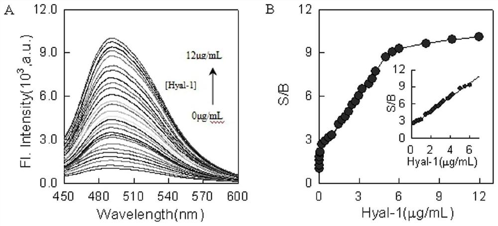
[0034] The determination of the titration curve was carried out under the optimized conditions. figure 2 (A) shows that after different concentrations of Hyal-1 (0-12 μg / mL), the fluorescence intensity of the nanosensor increases significantly with the increase of Hyal-1 concentration. and from figure 1 (B) It can be seen that after the nanosensor responds to Hyal-1, the size of the nanosensor after being degraded by Hyal-1 is about 5 nm. figure 2 (B) shows that the signal-to-background ratio (S / B) of the nanosensor and the analyte Hyal-1 in the range of 0.25-6.0 μg / mL exhibits a good linear relationship, and the linear function formula is y=1.267x+ 2.1811;R 2 =0.9956. The probe can accurately detect Hyal-1 in urine.
Embodiment 3
[0035] Example 3: Selective investigation of nanosensors
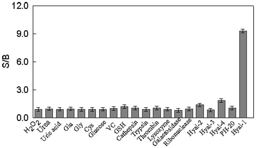
[0036] The nanosensor was added to PB buffer at a concentration of 0.02mmol / LTPC3@0.3mg / mL CHA, and then different representative interference species (0.1μM / mL H) were added to it. 2 O 2 ,Urea,Uric acid,Glu,Gly,Cys,Glucose,VC,GSH,Cathepsin,Trypsin,Thrombin,Galactosidase,Ribnudease,Hyal-2(5μg / mL),Hyal-3(5μg / mL),Hyal-4( 5 μg / mL), PH-20 (5 μg / mL) and Hyal-1 (5 μg / mL)). At the same time, add a blank group, only add 2 μL PBS buffer solution, shake well, measure and record the fluorescence intensity value of each group solution at excitation wavelength = 375 nm, emission wavelength = 495 nm, and obtain the effect of various species on the nano-sensing fluorescence signal. influences. The signal-to-background ratio is the fluorescence value of the solution after adding various interfering species divided by the fluorescence value of the blank group. The experiment was repeated three times and the average value was taken. ...
PUM
| Property | Measurement | Unit |
|---|---|---|
| particle diameter | aaaaa | aaaaa |
Abstract
Description
Claims
Application Information
 - R&D
- Intellectual Property
- Life Sciences
- Materials
- Tech Scout
- Unparalleled Data Quality
- Higher Quality Content
- 60% Fewer Hallucinations
Browse by: Latest US Patents, China's latest patents, Technical Efficacy Thesaurus, Application Domain, Technology Topic, Popular Technical Reports.
© 2025 PatSnap. All rights reserved.Legal|Privacy policy|Modern Slavery Act Transparency Statement|Sitemap|About US| Contact US: help@patsnap.com