Method, device and system for rapidly detecting bacterial drug resistance by utilizing nanopores
A nanopore, drug-resistant technology, applied in the field of bioengineering, can solve problems such as time-consuming, expensive, and small detection range
Pending Publication Date: 2021-01-29
WEST CHINA HOSPITAL SICHUAN UNIV
5 Cites 1 Cited by
AI-Extracted Technical Summary
Problems solved by technology
However, the detection of bacterial drug-resistant phenotypes requires sufficient time to culture Klebsiella pneumoniae, which is usually time-consuming; the detection of β-lactamase is fast, but th...
Method used
[0062] Carbapenem-resistant Klebsiella pneumoniae has rapidly become prevalent worldwide in recent decades, posing enormous challenges to today's clinical practice. Rapid detection of carbapenem-resistant Klebsiella pneumoniae could reduce inappropriate antimicrobial therapy and save lives. Traditional carbapenem-resistant Klebsiella pneumoniae detection methods are very time-consuming, and PCR and other sequencing methods are too expensive and technically demanding to meet clinical needs. Nanopore detection has the advantages of high sensitivity, real-time operation and low cost, and has been applied to the screening of disease biomarkers. In this study, we detected the amount of 16S rRNA in nucleic acid extracts after short-term culture of bacteria with the antibiotic imipenem to reflect the growth of bacteria to distinguish carbapenem-sensitive Klebsiella pneumoniae from Carbapenem-resistant Klebsiella pneumoniae. The specific signal generated after the probe binds to 16SrRNA can be recorded by using the MspA nanopore, so as to complete the ultrasensitive and rapid quantitative detection of 16S rRNA. We demonstrate that the nanopore assay can differentiate carbapenem-resistant Klebsiella pneumoniae from carbapenem-susceptible Klebsiella pneumoniae with an incubation time of only 4 hours. The time cost of this method is about 5% of that of the paper diffusion method, and the accuracy is close to that of the paper diffusion method. This new method has potential application value in rapid drug resistance detection of clinical microorganisms.
[0063] Specifically, nanopore sensing technology contributes to its wide application in third-generation DNA single-molecule sequencing. The nanometer-...
Abstract
The invention relates to a method and a device for detecting bacterial drug resistance and application of the method and the device. The method and the device are characterized in that bacterial growth is detected by utilizing a specific signal of a compound generated after a nanopore detection probe is combined with a bacterial biomarker and conducting quantitative detection on the bacterial biomarker. Compared with the prior art, the method and the device are high in detection sensitivity and high in speed, and have potential application value in the aspect of rapid drug resistance detectionof clinical microorganisms.
Application Domain
MicroorganismsMicrobiological testing/measurement +2
Technology Topic
NanoporeBacterial growth +6
Image
Examples
- Experimental program(3)
Example Embodiment
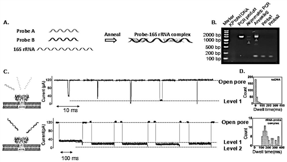
[0070] Example 1 Detection of 16S rRNA-probe complex
[0071] 1. Preparation of Bacterial Extracts
[0072] Two groups of Klebsiella pneumoniae samples from clinical patients were provided by West China Hospital of Sichuan University. Klebsiella pneumoniae samples were cultured to two different concentrations, the first group had a concentration of 0.5 MCF and the second group had a concentration of 4 MCF. At the beginning of the culture, the final concentration of imipenem used in the two groups was 16 mg/L, and the total RNA of Klebsiella pneumoniae was extracted by the TRIZOL method. First, collect 100 µL of the bacterial solution. The supernatant was removed after centrifugation (8000g, 4°C, 2 minutes). Precipitate with lysozyme and incubate at 37 °C for 10 min. Klebsiella pneumoniae were lysed, total RNA was extracted and washed with ethanol. Remove the centrifuge tube cap, dry at room temperature for 5-10min, add DEPC water or dissolve in rnas-free water. Add the RNase inhibitor to the dissolved solution to a final concentration of 20 U/μL for storage.
[0073] 2. Design probes and incubate with samples
[0074] The probes were designed to bind to specific segments of 16S rRNA so that the team of inventors could identify specific signals on target nucleic acids through the nanopore. Since the target 16S rRNA is 932bp long, it is difficult to distinguish the 16S rRNA-probe complex without a probe or a single probe, so the inventor team designed two probes to bind the specific expression of Klebsiella pneumoniae 16S rRNA. The two probes are the nucleotide sequences of probes A and B as shown in SEQ ID NO.1 and SEQ ID NO.2. The probes A and B were annealed with stored samples, and the formation of the probe 16S rRNA-probe complex was verified using agarose gel electrophoresis ( figure 2 in A).
[0075] 3. Results
[0076] The results of agarose gel electrophoresis showed that the 16S rRNA-probe complex ( figure 2 in B). In the sample of carbapenem-resistant Klebsiella pneumoniae, the residence time of the translocation signal of 16S rRNA-probe complex was in the range of 100-400ms, with a peak value of 196.98ms, and the residence time of single-stranded DNA translocation was between 0- In the range of 100ms, the peak value is 12.03ms ( figure 2 C and D in ). The dwell time of probe A and probe B is in the range of 0-70ms ( image 3 ). These results suggest that the long retention time signal is caused by the 16S rRNA-probe complex.
Example Embodiment
[0077] The optimization of embodiment two bacterial concentration and standard sample test
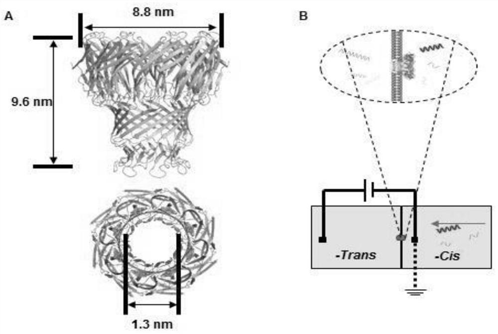
[0078] 1. Expression and purification of MspA nanopore
[0079] The gene of the MspA nanopore was cloned into the pET-28b plasmid, and the pET-28b plasmid carrying the MspA gene was transferred into the competent cells of the engineering bacteria BL21 Escherichia coli. At a temperature of 37°C, the successfully transferred Escherichia coli was cultured with LB medium, and kanamycin was added to 50 μg/ml. When the optical density (600nm) was close to 0.8, 0.8mM IPTG was added into the LB (lysogenic fermentation broth) medium, and the induction temperature was 15°C. After 12 hours of induction, E. coli were collected by centrifugation. The supernatant was collected after crushing E. coli with an ultrasonic generator, and further purified with an anion exchange column (Q-Sepharose) and molecular sieves (Superdex200 16/90). Purified proteins were analyzed by 10% SDS-PAGE (sodium dodecyl sulfate polyacrylamide gel electrophoresis). Purified MspA nanoporin can be aliquoted and stored at -80°C. Aliquoted samples are stable for years, and the nanopores retain their structural integrity when thawed.
[0080] 2. Nanopore electrophysiological signal detection experiment to determine the best sample concentration and the best bacterial culture time
[0081] 2.1 Determine the optimal sample concentration
[0082] A nanopore electrophysiological signal detection experiment was performed on the two bacterial extract samples with different concentrations in Example 1. The experimental method is:
[0083] The experiments were performed in a chamber provided by Warner Instruments. Nanopore electrophysiological signal detection experiments were performed at a voltage of 150 mV. The conductive buffer solution for the cis-side and trans-side is 400 mM KCl solution containing 10 mM HEPES, pH 7.0. A bilayer lipid membrane (BLM) coated on both sides of a 150 μm pore was formed from 1,2-dihydroxyformyl-sn-glycero-3-phosphocholine (DPHPC). Addition of MspA to the solution in the cis compartment allows insertion of the MspA protein and faster formation of the BLM. A single MspA nanopore embedment will result in an increased current, corresponding to a conductance of 1.2 nS. After recording current signals inserted into a single MspA nanopore through a Heka EPC-10 patch clamp (HEKA), samples were added to the cis side.
[0084] Two concentrations of K. pneumoniae were used to optimize detection efficiency. In the sample of 0.5MCF, the target RNA transfer signal frequency of the control group was 0.02±0.02/min (n=3), and the target RNA transfer signal frequency of the carbapenem-resistant Klebsiella pneumoniae group was 0.13±0.05/min. min (n=3). While in the 4MCF samples, the target RNA translocation frequency in the control group was 0 per minute (n=3), and the translocation frequency in the carbapenem-resistant Klebsiella pneumoniae group was 0.33±0.07 per minute (n= 3)( Figure 4 in A). The 4MCF sample was better detected in the nanopore assay compared to the 0.5MCF sample.
[0085] 2.2. Determine the best bacterial culture time
[0086] Total RNA extracted from carbapenem-resistant Klebsiella pneumoniae and carbapenem-susceptible Klebsiella pneumoniae was incubated with probe A and probe B, and the post-incubation RNA was detected by MspA nanopore, respectively. solution ( Figure 4 in B). Two parameters of the signal obtained by measuring the sample through the nanopore, blocking rate and residence time are plotted into a scatter plot ( Figure 4 In C), significant differences in residence time between the different groups can be observed, especially in the range of blockage ratio 0.6 to 0.8 and residence time 100 msec to 400 msec. Therefore, the signal within this range is selected as the specific signal for diagnosis. After comparing the number of 16SrRNA-probe signals in a given range from blank, control, carbapenem-resistant Klebsiella pneumoniae and carbapenem-sensitive Klebsiella pneumoniae samples, f= 0.1 min -1 Target signal translocation frequency thresholds to differentiate carbapenem resistance in Klebsiella pneumoniae. To determine the minimum bacterial incubation time required to distinguish carbapenem-resistant Klebsiella pneumoniae from carbapenem-sensitive Klebsiella pneumoniae, samples with different bacterial incubation times, including 2 Hours, 4 hours and 8 hours, the experimental results show that 4 hours is the best bacterial culture time taking into account both sensitivity and efficiency.
Example Embodiment
[0087] Example 3 Double-blind test of MspA nanopore detection of clinical samples
[0088] Bacteria in blood samples from 20 patients with Klebsiella pneumoniae infection provided by West China Hospital were cultured, total RNA was extracted and used for double-blind experiments. Each sample was assayed at least three times with the MspA nanopore. After analysis, the number of 16S rRNA probe signals with a blocking rate of 0.6 to 0.8 and a residence time of 100 ms to 400 ms was collected and compared with the target signal translocation frequency threshold fthreshold.
[0089] Among the 20 samples, as shown in Table 1, 9 of them are above the threshold (0.1 min -1 ) and was judged to be carbapenem-resistant Klebsiella pneumoniae. As shown in Table 2, the other 11 samples are below the threshold of 0.1 min -1 , these clinical samples were determined to be carbapenem-sensitive Klebsiella pneumoniae samples ( Figure 5 in A). Compared with assay results obtained from standard clinical methods (disk diffusion method or PCR), the nanopore assay method of the present invention has the advantages of low cost and short time consumption (Table 3). The results of 18 samples measured by nanopore are correct ( Figure 5 In B), there are two false negative results.
[0090] Table 1. Information on clinical samples of carbapenem-susceptible Klebsiella pneumoniae
[0091]
[0092]
[0093] Note: the sample ID is the patient ID in the hospital, and the sample number is the corresponding number in the study of the present invention.
[0094] Table 2. Information on clinical samples of carbapenem-resistant Klebsiella pneumoniae
[0095] Sample ID sample# SCIM(mm) drug resistance gene drug resistance gene 17012889-3 1 6 KPC KPC-2 17019349-3 3 6 KPC KPC-2 1810143046 4 6 KPC KPC-2 15043287-1 5 6 KPC KPC-2 15057156-1 6 6 KPC KPC-2 15083593-1 7 6 KPC KPC-2 1807191036 8 6 KPC KPC-2 1807271015 9 24 burden - 17008404-1 11 6 KPC No 17012837-3 12 6 KPC KPC-2 17020362-3 20 6 KPC KPC-2
[0096] Note: the sample ID is the patient ID in the hospital, and the sample number is the corresponding number in the study of the present invention.
[0097] Table 3. Comparison of different detection methods for carbapenem-resistant Klebsiella pneumoniae
[0098]
[0099] The above examples use the software Clampfit 10.6 and Origin Pro 8.0 for data analysis. Blocking current is defined as ΔI/I 0 , where I 0 is the current for a fully open pore, and ΔI is the amplitude of the blocking current induced by the transport molecule. Dwell times were collected by the single-channel search function of Clampfit 10.6. These two parameters were used to quantify target 16S rRNA from surviving carbapenem-resistant Klebsiella pneumoniae. All data were obtained from 20-min electrophysiological recordings, and the experimental groups were repeated 3 times independently.
[0100] Technical problem and solution summary of the present invention
[0101] Rapid and accurate detection of carbapenem resistance in Klebsiella pneumoniae is very important in clinical treatment. However, the current detection technology cannot fully meet the clinical needs. Our goal is to develop a novel detection method for carbapenem resistance of Klebsiella pneumoniae based on nanopore sensing technology to solve the clinical problems.
[0102] In the past 20 years, protein nanopore expression, purification and electrophysiological detection technologies have developed rapidly, and nucleic acid detection methods based on various types of protein nanopores have been very mature. In the present invention, the inventor team designed two DNA probes to specifically bind the 16S rRNA of Klebsiella pneumoniae with carbapenem resistance, and the translocation of the 16S rRNA-probe complex through the MspA nanopore will cause Dwell time between 100ms and 400ms. According to the blocking rate and retention time of the specific blocking signal, 16S rRNA in carbapenem-resistant Klebsiella pneumoniae samples could be detected ( Image 6 ). The method can be used to distinguish carbapenem-resistant Klebsiella pneumoniae standard samples from carbapenem-resistant and carbapenem-sensitive Klebsiella pneumoniae Klebsiella pneumoniae samples sensitive to carbapenems, and the incubation time of bacterial samples is only 4 hours.
[0103] In addition, the inventor team used MspA nanopores to measure 20 clinical samples provided by West China Hospital. Among the 11 clinical samples of carbapenem-resistant Klebsiella pneumoniae, 9 samples were correctly diagnosed and 2 samples were detected as false negative; in 9 cases of carbapenem-sensitive pneumonia Among the Klebsiella samples, all 9 samples were correctly diagnosed. The nanopore diagnostic method was 90 percent accurate. RNA degradation during sample storage or transfer is the leading cause of 10% of false-negative diagnoses. The transportation process of clinical samples from the hospital to the laboratory and the time interval between sample processing and nanopore assay increase the possibility of RNA degradation, resulting in reduced amounts of 16S rRNA and specific blocking signals.
[0104] The above results are based on the experimental research of the new nanopore single-molecule diagnostic technology. Further verification research aimed at improving the sensitivity can be carried out around the following aspects: optimization of bacterial culture conditions and RNA extraction technology; detection of 16SrRNA-probe complexes Modification and transformation of protein nanopores; testing with a large number of clinical samples and improving statistical methods for data processing. Stable storage of aliquoted MspA protein at -80°C enabled mass production of nanopores for subsequent multiple testing.
[0105] In summary, the research of the inventor team confirmed that the nanopore single-molecule detection technology can be used for rapid clinical diagnosis of carbapenem-resistant Klebsiella pneumoniae. Compared with the disc diffusion method or PCR method, the two most widely used methods in clinical diagnosis, the nanopore detection method has the advantages of low cost, high efficiency and easy operation. This method can be used in clinical laboratory diagnosis as a supplement to existing diagnostic methods. With the development of nanopore chip technology, the detection of multiple clinical samples based on nanopore arrays will be further applied to clinical point-of-care diagnosis.
PUM


Description & Claims & Application Information
We can also present the details of the Description, Claims and Application information to help users get a comprehensive understanding of the technical details of the patent, such as background art, summary of invention, brief description of drawings, description of embodiments, and other original content. On the other hand, users can also determine the specific scope of protection of the technology through the list of claims; as well as understand the changes in the life cycle of the technology with the presentation of the patent timeline. Login to view more.