
Artificial ovarian stent constructed by utilizing 3D biological printing technology, artificial ovary, construction method and application

A 3D printing and bioprinting technology, applied in the field of artificial ovaries, can solve the problems of difficult long-term simulation of ovaries, insufficient mechanical strength, easy degradation, etc., and achieve the effects of strong repeatability, improved maturity rate, and good stability

Pending Publication Date: 2021-02-23
TONGJI HOSPITAL ATTACHED TO TONGJI MEDICAL COLLEGE HUAZHONG SCI TECH
View PDF4 Cites 0 Cited by
  • Summary
  • Abstract
  • Description
  • Claims
  • Application Information

AI Technical Summary

Problems solved by technology

The materials used to construct artificial ovaries are mostly natural sources, including collagen, fibrinogen, sodium alginate and hyaluronic acid, etc., but they often have insufficient mechanical strength and are easily degraded in the body, making it difficult to simulate the function of the ovary for a long time

Method used

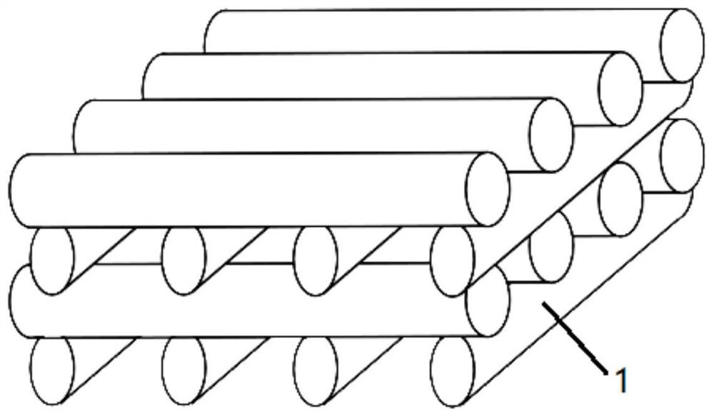

Image

  • Artificial ovarian stent constructed by utilizing 3D biological printing technology, artificial ovary, construction method and application
  • Artificial ovarian stent constructed by utilizing 3D biological printing technology, artificial ovary, construction method and application
  • Artificial ovarian stent constructed by utilizing 3D biological printing technology, artificial ovary, construction method and application

Examples


Embodiment 1

[0037] In this example, methacryloyl hydrogel was used to construct a mouse artificial ovary scaffold to promote the maturation of immature oocytes.

[0038] The granulosa cells isolated from the mouse ovary were added to the cell culture plate for culture, and the culture medium was McCoy's 5A medium, supplemented with 10% fetal bovine serum, 10ng / ml androgen and 0.1% penicillin-streptomycin mixture by volume, The percentages of each component are volume percentages after addition, and the concentration is the concentration in the culture medium after addition; put the culture plate at 37°C with a volume fraction of 5% CO 2 Cultured in a cell incubator, the cells in the logarithmic growth phase were digested with 0.25% (mass volume ratio, equivalent to 0.25g trypsin dissolved in 100ml Hank's balanced salt solution) trypsin to obtain a granular cell suspension.

[0039] The methacrylylated hydrogel (concentration is 20%), the resuspended granule cells and the blue light initia...

Embodiment 2

[0046] In this example, methacryl hydrogel was used to construct a human artificial ovary scaffold to promote the maturation of immature oocytes.

[0047] Human granulosa cells are derived from women who received in vitro fertilization-embryo transfer or cytoplasmic sperm microinjection in the reproductive center of the hospital. The follicular fluid obtained on the day of egg collection was recovered, and the follicular fluid was centrifuged at 500r / min for 5min, and the supernatant was discarded. Resuspend the pellet with phosphate buffered saline (PBS), then add an equal volume of human peripheral blood lymphocyte separation medium (Ficoll separation medium), centrifuge at 2000r / min for 20min, obtain ovarian granulosa cell mucus mass, and extract the middle layer mucus mass Resuspend in the culture medium and pipette repeatedly to obtain the original suspension containing granule cells.

[0048] Granulosa cells isolated from human follicular fluid were added to cell culture...


PUM


Abstract

The invention discloses an artificial ovarian stent constructed by using a 3D biological printing technology. The artificial ovarian stent is obtained by performing 3D printing and curing with biological ink which is mixed with granular cells and is used for 3D printing. The granular cells refer to rhombus or flat cells which surround oocytes in follicles in the follicle phase of ovaries of mammals, and the cytoplasm of the granular cells contains granules. The invention further provides a construction method of the artificial ovarian stent, an artificial ovary constructed by the artificial ovarian stent as well as a construction method and application of the artificial ovary. In the 3D printing process, the ovarian granular cells are creatively added into the biological ink, so that the granular cells in the whole stent are uniformly distributed, more growth signals are provided for growth of follicles on the stent, and the maturation rate of oocytes is increased.

Description

technical field [0001] The present invention relates to an artificial ovary, in particular to an artificial ovary scaffold constructed by 3D bioprinting technology, an artificial ovary and its construction method and application. Background technique [0002] Worldwide, tumors remain a major problem threatening life and health. In recent years, with the advancement of diagnostic techniques and treatment methods, the 5-year survival rates of adults and children have reached 65% and 83%, respectively. However, due to the side effects of radiotherapy and chemotherapy, nearly 1 / 3 of women will lose their fertility as a result. In addition, women with premature ovarian insufficiency (POI) also face the same problem. They lose ovarian function before the age of 40 and experience symptoms related to menopause. To help these women, more and more research has begun to focus on how to preserve their fertility. This is not only a medical treatment that can improve the quality of lif...

Claims


Application Information

Patent Timeline
IPC IPC(8): A61L27/38A61L27/24A61L27/52B33Y70/10C12N5/075
CPCA61L27/3804A61L27/24A61L27/52C12N5/0609B33Y70/10C12N2509/00C12N2501/392C12N2500/25C12N2501/31C12N2501/11C08L89/00
Inventor 王世宣张金金吴桐高越越
Owner TONGJI HOSPITAL ATTACHED TO TONGJI MEDICAL COLLEGE HUAZHONG SCI TECH
Learn More