Gene editing microneedle for treating inflammatory skin diseases and application thereof

An inflammatory skin disease, gene editing technology, applied in skin diseases, microneedling, gene therapy, etc., can solve problems such as difficulty in reaching the epidermis and dermis

Active Publication Date: 2021-05-18
ZHEJIANG UNIV
View PDF5 Cites 2 Cited by
  • Summary
  • Abstract
  • Description
  • Claims
  • Application Information

AI Technical Summary

Problems solved by technology

In addition, although these small molecules have a good antagonistic effect on NLRP3, in inflammatory skin diseases, the activation of NLRP3 inflammasome is mainly located in the epidermis and dermis of human skin, and it is difficult for these inhibitors to reach the epidermis and dermis when administered orally. Dermis

Method used


Image

  • Gene editing microneedle for treating inflammatory skin diseases and application thereof
  • Gene editing microneedle for treating inflammatory skin diseases and application thereof
  • Gene editing microneedle for treating inflammatory skin diseases and application thereof

Examples


Embodiment 1

[0023] Example 1: Preparation of gene-edited microneedle patch

[0024] This embodiment includes the following raw materials by mass percentage:

[0025] Needle tip matrix: collagen tripeptide 10%, sodium hyaluronate (molecular weight 34kDa) 10%, the rest is water.

[0026] In addition, every 10 mL of sodium hyaluronate + collagen tripeptide solution in the microneedle tip matrix contains 60 μg of dexamethasone, 20 μg of Cas9 protein, and 5 μg of sgRNA.

[0027] Base matrix: collagen tripeptide 10%, sodium hyaluronate (molecular weight 34kDa) 10%, the rest is water.

[0028] The preparation method of this embodiment:

[0029] 1. According to the prescription ratio, take 1g sodium hyaluronate (molecular weight 34kDa) and 1g collagen tripeptide and disperse in 10mL water, stir until fully dissolved, add the dexamethasone nanocomposite wrapped in degradable polymer carrier in the prescription ratio PLGA / Dex and degradable cationic carrier wrap the gene editing ribonucleoprotei...

Embodiment 2

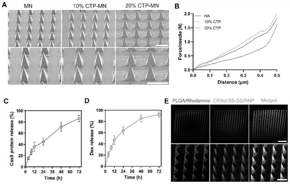
[0031] Example 2: Characterization of gene-edited microneedle patches

[0032] The specific operation method is as follows: use a scanning electron microscope to observe the morphology of microneedles with different percentages of collagen tripeptides. Place the microneedles containing different percentages of collagen tripeptides on the texture analyzer with the needle point facing up, and the probe of the texture analyzer moves down at a speed of 1mm / s, and the mechanical strength is reflected by measuring the curve of displacement and force.

[0033] In order to investigate the dissolution of the microneedle in the skin, the double-loaded microneedle patch was applied to the back skin of the mouse for 0, 1, 3 and 5 minutes, then removed, and then the remaining microneedle patch was taken out and photographed with a scanning electron microscope.

[0034] In order to evaluate the drug release behavior of the microneedles in vitro, the double-loaded microneedle patch was first...

Embodiment 3

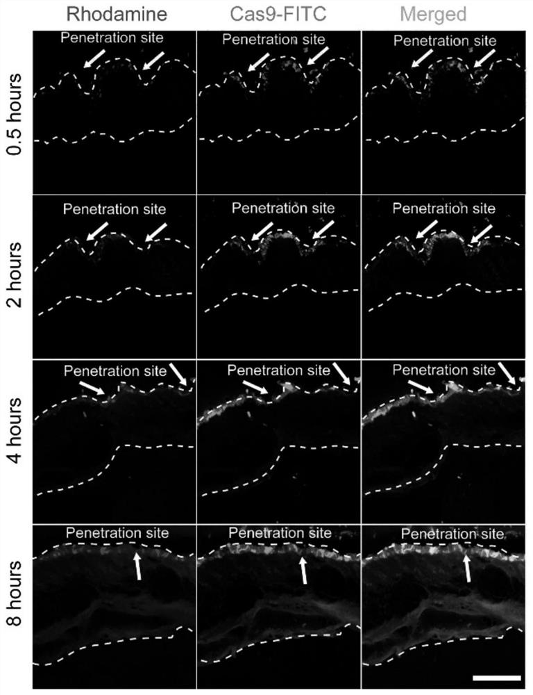
[0036] Example 3: Drug distribution after the gene-edited microneedle patch pierces the skin

[0037] In order to study the degradation behavior of microneedles in vivo, a double-loaded microneedle patch loaded with PLGA / rhodamine B and CP / Ad-SS-GD / Cas9protein-FITC was prepared, and then the double-loaded microneedle patch was applied to the back of the mouse The back skin tissue was separated from the skin at different times for frozen embedding, and then the tissue was placed in a frozen section for sectioning, and the fluorescence distribution was observed under a laser confocal microscope

[0038] Experimental results: if figure 2 As shown, the microneedle patch can be successfully penetrated into the mouse skin with a depth of about 300 μm. After piercing into the mouse skin, the tip of the microneedle can quickly absorb the liquid in the skin tissue and dissolve rapidly. At the same time, rhodamine and Cas9-FITC Released in large quantities, distributed in the epidermi...


PUM

PropertyMeasurementUnit
molecular weightaaaaaaaaaa
diameteraaaaaaaaaa

Abstract

The invention provides a gene editing microneedle for treating inflammatory skin diseases and application thereof. The gene editing microneedle comprises a needle tip part and a base part, wherein the needle tip part comprises two nano compounds and a needle tip matrix, and the base part is a base matrix. The gene editing microneedle can improve the gene editing efficiency, deliver the retention of glucocorticoid drugs and gene editing ribonucleoprotein compounds in the skin, and synergistically improve the skin inflammation environment. The microneedle has the skin barrier function of repairing skin lesion tissues, thereby achieving the inflammatory process of relieving psoriasis and atopic dermatitis. The invention also provides the application of the gene editing microneedle in the preparation of drugs for treating inflammatory skin diseases.

Description

technical field [0001] The invention relates to the fields of pharmacy, biotechnology, polymer chemistry, cell biology, etc., and specifically relates to a gene editing microneedle for treating inflammatory skin diseases and its application. It is a new type of microneedle patch to deliver gene-edited ribonucleoprotein complexes, as well as the application of drugs for the preparation of inflammatory skin diseases. Background technique [0002] Inflammatory skin diseases (ISDs) are a group of common diseases in life, and their pathological features are the activation of innate and adaptive immune responses through the production of pro-inflammatory cytokines. Among various inflammatory skin diseases, psoriasis and atopic dermatitis are increasingly becoming the main problems that threaten public health, and the course of the disease lasts for a long time, making it difficult to be completely cured. Psoriasis is an immune-mediated inflammatory skin disease characterized clin...

Claims


Application Information

Patent Timeline
Patent Type & Authority Applications(China)
IPC IPC(8): A61K9/00A61K31/573A61K48/00A61K9/51A61K47/36A61K47/42A61K47/69A61P17/06A61P17/00A61M37/00
CPCA61K9/0021A61K31/573A61K9/5153A61K47/6951A61K47/36A61K47/42A61K48/005A61P17/00A61P17/06A61M37/0015A61M2037/0053
Inventor 平渊潘齐万涛
Owner ZHEJIANG UNIV
Why Eureka
Try Eureka