Preparation method of Se-coated BSA nano-drug and application of Se-coated BSA nano-drug in acute kidney injury

A technology for acute kidney injury and nano-drugs, which can be used in drug combinations, non-active components of polymer compounds, active components of sulfur/selenium/tellurium, etc. Toxicity, slow progression, prevent assembly

Active Publication Date: 2021-07-09
THE SECOND AFFILIATED HOSPITAL ARMY MEDICAL UNIV
View PDF6 Cites 1 Cited by
  • Summary
  • Abstract
  • Description
  • Claims
  • Application Information

AI Technical Summary

Problems solved by technology

However, the specific mechanism of selenium in acute kidney injury is still uncl

Method used


Image

  • Preparation method of Se-coated BSA nano-drug and application of Se-coated BSA nano-drug in acute kidney injury
  • Preparation method of Se-coated BSA nano-drug and application of Se-coated BSA nano-drug in acute kidney injury
  • Preparation method of Se-coated BSA nano-drug and application of Se-coated BSA nano-drug in acute kidney injury

Examples


Embodiment 1

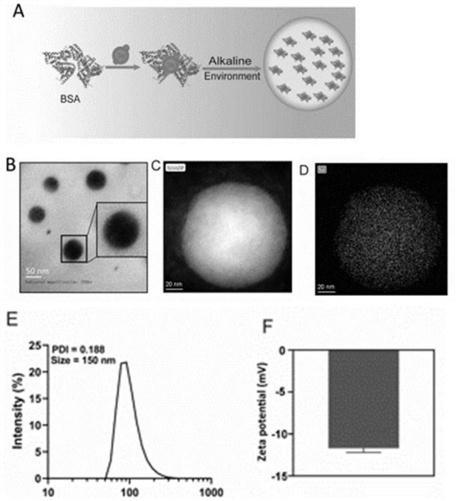
[0026] Preparation and analysis of physicochemical properties of Se@BSA nanomedicine

[0027] Dissolve 40mg BSA in 10mL water and stir for 1h, add 10mL 0.05M ascorbic acid solution to react for 0.5h, then add 4mL 0.05M Na 2 SeO 3 The solution was reacted for 1 hour, reacted for 3 hours, adjusted to pH 8, reacted for 10 hours, dialyzed with 14000 dialysis bag for 2 days, and freeze-dried to obtain Se@BSA nanomedicine. The prepared Se@BSA nanomedicine was detected by transmission electron microscopy (TEM).

[0028] The result is as figure 1 As shown in A-F, the Se@BSA nanomedicine is spherical and uniform in size. The particle size of the Se@BAS nanomedicine is about 150nm, and the surface charge is about -12mv.

Embodiment 2

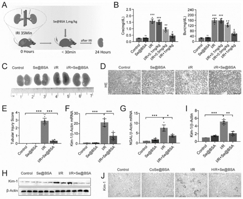
[0030] The role of Se@BSA nanomedicine in ischemia / reperfusion-induced acute kidney injury

[0031] A total of 40 C57BL / 6 mice with a body weight of 20-22 g (Beijing HFK Biotechnology Company) were selected, and the mice were randomly (n=10) divided into four groups: sham operation, sham operation+Se@BAS, I / R and I / R+Se@BAS.

[0032] Such as figure 2 As shown in the schematic diagram of the ischemia / reperfusion operation in middle A, the bilateral kidneys of mice in the I / R group were clipped for 35 minutes; in the I / R+Se@BAS group, different doses (0, 0.1 , 0.5, 1mg / kg) of Se@BAS nanospheres; in the sham operation group, the same dose of PBS was injected; in the sham operation+Se@BAS (1mg / kg) group, the same dose of PBS and Se@BSA nanospheres were injected . Serum samples were collected and stored at -80°C, using creatinine (CRE) detection kit (Nanjing Jiancheng Bio, C011-2-1) and urea nitrogen (BUN) kit (Nanjing Jiancheng Biology, C013-2-1) Detect CRE and BUN. The resul...

Embodiment 3

[0049] Safety evaluation of Se@BSA nanomedicine

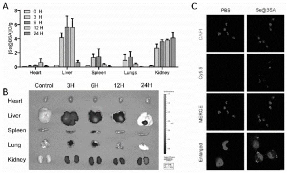
[0050] (1) Distribution of Se@BSA nanomedicine in mice

[0051] The mice were injected with Se@BSA (1mg / kg), and the mice were detected by in vivo imaging at 3, 6, 12h, and 24h. After 24h, the heart, liver, spleen, lung, and kidney tissues were taken, and selenium was determined by ICP-MS content. The result is as image 3 As shown in A and B, ICP-MS results show that the content of Se@BSA nanospheres in various organs increases with time, mainly enriched in liver and kidney; mouse imaging results show that Se@BSA nanospheres in The biodistribution in mice was time-dependent. Se@BSA was mainly concentrated in the liver and kidney, peaked at 6 hours after injection, and mainly concentrated in the kidney at 24 hours. The selenium content in the organs decreased significantly, but the Se concentration in the kidney increased at 24 hours, which was similar to the ICP-MS results, indicating that Se@BSA had partial kidney targetin...


PUM

PropertyMeasurementUnit
Particle sizeaaaaaaaaaa
Surface chargeaaaaaaaaaa

Abstract

The invention relates to a preparation method of a Se-coated BSA nano-drug and application of the Se-coated BSA nano-drug in acute kidney injury, and belongs to the field of biological materials. The Se-coated BSA nano-drug synthesized by the invention is a safe nano-material capable of being used for intravenous injection, can be used for managing injury caused by hypotension of ischemic kidney and cardiovascular surgery, and aims to allow Se-coated BSA to be released after kidney I/R so as to avoid systemic toxicity, and the specific mechanism is that Se-coated BSA up-regulates expression of Gpx1 mRNA and protein level, removes ROS, inhibits accumulation of NLRP3 to prevent assembly of inflammatory bodies. The activation of a downstream inflammation signaling pathway and the phosphorylation of NF-beta B are realized, and the progress from AKI to CKD is slowed down.

Description

technical field [0001] The invention belongs to the field of biological materials, and relates to a Se@BSA nano-medicine and its application in acute kidney injury. Background technique [0002] Acute kidney injury (AKI) is a critical illness with high morbidity and mortality, especially for patients admitted to the intensive care unit (ICU). AKI is mainly caused by sepsis, ischemia / reperfusion and nephrotoxin and is characterized by rapid loss of renal function, leading to accumulation of metabolic waste products, electrolyte and fluid imbalance. Ischemia / reperfusion injury (I / R) is AKI that causes tubular epithelial cells (TECs) to generate a large amount of reactive oxygen species (ROS), triggering mitochondrial damage and lipid peroxidation, resulting in destructive cellular damage. Inflammatory factors produced by TECs lead to the migration and infiltration of a large number of inflammatory cells, further exacerbating renal injury. Despite the rapid advances in basic ...

Claims


Application Information

Patent Timeline
IPC IPC(8): A61K33/04A61K47/42A61K9/14A61P13/12
CPCA61K33/04A61K9/146A61P13/12
Inventor 王少博赵景宏陈银王成黄英辉
Owner THE SECOND AFFILIATED HOSPITAL ARMY MEDICAL UNIV
Why Eureka
Try Eureka