Visual monodisperse embolism microsphere with internal radiotherapy performance and preparation method of visual monodisperse embolism microsphere
A technology for radiotherapy and embolization of microspheres, applied in the field of biomedicine, can solve the problems of invisible size, non-uniformity, non-degradability, etc., and achieve the effects of uniform shape, low production cost, and uniform and controllable size.
- Summary
- Abstract
- Description
- Claims
- Application Information
AI Technical Summary
Problems solved by technology
Method used
Image

Examples
Embodiment 1
[0047] In this example, visualized monodisperse embolic microspheres with internal radiotherapy properties ( 131 1-poly GelMA microspheres), the steps are as follows:
[0048] (1) Preparation of internal phase, external phase fluid and collection solution
[0049] Prepare the internal phase fluid: Add the photoinitiator phenyl 2,4,6-trimethylbenzoyl lithium phosphonate (LAP) into deionized water, dissolve it in a water bath at 50°C to obtain a photoinitiator solution, add formazan Acrylylated gelatin (GelMA, double bond substitution degree is 90%) was added to the photoinitiator solution, dissolved uniformly in a water bath at 50°C, and filtered to obtain the internal phase fluid; in the internal phase fluid, deionized water, GelMA, LAP The mass ratio is 1:0.05:0.0025.
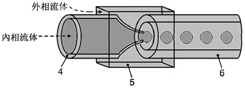
[0050] Prepare the external phase fluid: dissolving polyglycerin ricinoleate (PGPR) in soybean oil to obtain the external phase fluid; the mass ratio of soybean oil to PGPR in the external phase fluid is 1:0...
Embodiment 2
[0061] In this example, the preparation 131 I-poly GelMA microspheres, the steps are as follows:
[0062] (1) Preparation of internal phase, external phase fluid and collection solution
[0063] Prepare the internal phase fluid: add the photoinitiator LAP to deionized water, dissolve it uniformly in a water bath at 50°C to obtain a photoinitiator solution, add GelMA (the degree of substitution of the double bond is 90%) to the photoinitiator solution, and heat it at 50°C Dissolve evenly in a water bath, filter to obtain the internal phase fluid; the mass ratio of deionized water, GelMA, and LAP in the internal phase fluid is 1:0.05:0.0025.
[0064] Prepare the external phase fluid: dissolve PGPR in soybean oil to obtain the external phase fluid; the mass ratio of soybean oil to PGPR in the external phase fluid is 1:0.05.
[0065] Prepare the collection solution: the collection solution is the same as the external phase fluid.
[0066] (2) Preparation of monodisperse poly Ge...
Embodiment 3
[0075] In this example, the preparation 131 I-poly GelMA microspheres, the steps are as follows:
[0076] (1) Preparation of internal phase, external phase fluid and collection solution
[0077] Prepare the internal phase fluid: add the photoinitiator LAP to deionized water, dissolve it uniformly in a water bath at 50°C to obtain a photoinitiator solution, add GelMA (the degree of substitution of the double bond is 90%) to the photoinitiator solution, and heat it at 50°C Dissolve evenly in a water bath, filter to obtain the internal phase fluid; the mass ratio of deionized water, GelMA, and LAP in the internal phase fluid is 1:0.1:0.0025.
[0078] Prepare the external phase fluid: dissolve PGPR in soybean oil to obtain the external phase fluid; the mass ratio of soybean oil to PGPR in the external phase fluid is 1:0.05.
[0079] Prepare the collection solution: the collection solution is the same as the external phase fluid.
[0080] (2) Preparation of monodisperse poly Gel...
PUM
| Property | Measurement | Unit |
|---|---|---|
| Particle size | aaaaa | aaaaa |
| Concentration | aaaaa | aaaaa |
| Particle size | aaaaa | aaaaa |
Abstract
Description
Claims
Application Information
 - R&D
- Intellectual Property
- Life Sciences
- Materials
- Tech Scout
- Unparalleled Data Quality
- Higher Quality Content
- 60% Fewer Hallucinations
Browse by: Latest US Patents, China's latest patents, Technical Efficacy Thesaurus, Application Domain, Technology Topic, Popular Technical Reports.
© 2025 PatSnap. All rights reserved.Legal|Privacy policy|Modern Slavery Act Transparency Statement|Sitemap|About US| Contact US: help@patsnap.com