Preparation and application of a black phosphorus nanosheet-supported indocyanine green nanosystem

An indocyanine green and nanosheet technology, which is applied in the field of clinical imaging of diagnostic agents, can solve the problems of reducing fluorescence imaging effect and poor stability, and achieves the effects of simple and easy operation, mild preparation conditions, and reduced toxic and side effects.

Active Publication Date: 2022-07-15
UNIV OF SCI & TECH OF CHINA
View PDF3 Cites 0 Cited by
  • Summary
  • Abstract
  • Description
  • Claims
  • Application Information

AI Technical Summary

Problems solved by technology

However, indocyanine green has poor stability in polar dispersions, and it is easy to aggregate to form polymers, thereby reducing the effect of fluorescence imaging. Second, the polymers are quickly cleared in plasma

Method used


Image

  • Preparation and application of a black phosphorus nanosheet-supported indocyanine green nanosystem
  • Preparation and application of a black phosphorus nanosheet-supported indocyanine green nanosystem
  • Preparation and application of a black phosphorus nanosheet-supported indocyanine green nanosystem

Examples


Embodiment 1

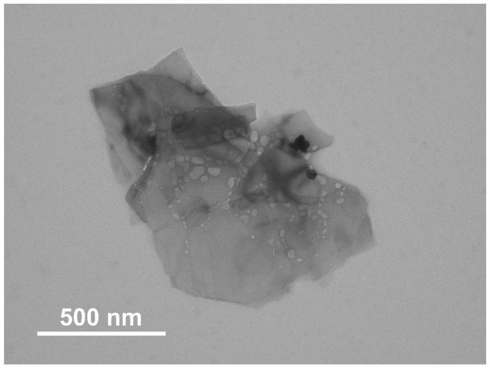
[0040] 1. Pour nitrogen into the stripped black phosphorus suspension and keep it in this state for 30 minutes to remove oxygen. Use a domestic high-speed centrifuge at 2500 rpm to screen out the black phosphorus nanosheet precipitate of this size. The obtained black phosphorus nanosheets were dispersed in ethanol, and the particle size was determined by transmission electron microscope. The result is as figure 1 As shown, the prepared black phosphorus nanosheets have a size of 892 ± 16 nm

[0041] 2. Weigh 25 mg of the obtained black phosphorus nanosheets, disperse it into 25 mL of normal saline, and place it in a 50 mL Shrek bottle. Nitrogen gas was introduced into it, kept in this state for 30 minutes, and sealed with a rubber stopper. It was then placed in a water bath at 25°C for 2 hours to sonicate, so that the black phosphorus nanosheets were uniformly dispersed. Then, 2.5 mL of indocyanine green (10 mg / mL) dispersion was slowly poured into it, and 20 mL of aminated...

Embodiment 2

[0043] 1. Pour nitrogen into the stripped black phosphorus suspension and keep it in this state for 30 minutes to remove oxygen. Using a domestic high-speed centrifuge at 7500 rpm, the black phosphorus nanosheet precipitate of this size was screened out. The obtained black phosphorus nanosheets were dispersed in ethanol, and the particle size was determined by transmission electron microscope. The result is as image 3 As shown, the size of the prepared black phosphorus nanosheets is 312±12 nm.

[0044] 2. Weigh 25 mg of the prepared black phosphorus nanosheets, disperse it into 25 mL of normal saline, and place it in a 50 mL Shrek bottle. Nitrogen gas was introduced into it, kept in this state for 30 minutes, and sealed with a rubber stopper. It was then placed in a water bath at 25°C for 2 hours to sonicate, so that the black phosphorus nanosheets were uniformly dispersed. Then, 2.5 mL of indocyanine green (10 mg / mL) dispersion was slowly poured into it, and 20 mL of ami...

Embodiment 3

[0046] Weigh a certain amount of ICG@BPNS-PEG solid and free indocyanine green screened at different speeds, and the concentration of indocyanine green was kept at 2 mg / mL. Three groups of female BALB / c nude mice (4-6 weeks, body weight 15-20 mg) were injected subcutaneously with 2*10 5 4T1 breast cancer cells, when the tumor volume reaches 50mm 3 time (tumor volume calculation formula: tumor length × (tumor width) 2 / 2), two groups of mice were injected with ICG@BPNS-PEG at different rotational speeds into the tail vein as the experimental group, and the remaining group of mice were injected with free indocyanine green into the tail vein as the control group. Take pictures and observe under the small animal imager after 6 hours,

[0047] Compared figure 1 and Figure 4 As a result, the size of the black phosphorus nanosheets screened by high-speed centrifugation is smaller, and the black phosphorus nanosheets also have the structural characteristics of thin layers. The sm...


PUM

PropertyMeasurementUnit
sizeaaaaaaaaaa

Abstract

The invention discloses the preparation and application of a black phosphorus nanosheet-loaded indocyanine green nanometer system. Black phosphorus nanosheets of different sizes are used as carriers, which have a large specific surface area and can effectively realize high loading of indocyanine green; In addition, through the modification of the aminated polyethylene glycol in the present invention, the dispersion stability of the constructed nanomaterial in the polar dispersion liquid is greatly improved. The black phosphorus nanosheet-loaded indocyanine green nanometer system prepared by the invention can be used as a fluorescent imaging agent, can effectively realize tumor tissue imaging, can distinguish the tumor boundary position, and enhance the fluorescent imaging effect of the tumor site.

Description

technical field [0001] The invention belongs to the technical field of clinical imaging of diagnostic agents, in particular to the preparation and application of a black phosphorus nanosheet-loaded indocyanine green nanometer system. Background technique [0002] Fluorescence imaging technology is an imaging technology based on injecting fluorescent molecules into animals or humans and detecting emitted light waves to visualize tissues or organs. As a complementary clinical imaging technology, fluorescence imaging has the advantages of: first, it does not use any dangerous ionization or radiation; second, the imaging instrument is cheap, easy to operate, and has a short sampling time, enabling real-time intraoperative imaging. [0003] Indocyanine green, also known as ICG, is a trihydrocarbon dye with a maximum absorption wavelength and emission wavelength of 740nm and 800nm. It has the advantage of strong tissue penetration. It has been approved by the US Food and Drug Admi...

Claims


Application Information

Patent Timeline
Patent Type & Authority Patents(China)
IPC IPC(8): A61K49/00B82Y5/00
CPCA61K49/0034A61K49/005A61K49/0054B82Y5/00
Inventor 胡源马小鹏陈韦剑潘婉婉
Owner UNIV OF SCI & TECH OF CHINA
Why Eureka
Try Eureka