Solid nanopore-fluorescence resonance energy transfer composite detection method and system
A fluorescence resonance energy and composite detection technology, applied in fluorescence/phosphorescence, measurement devices, material electrochemical variables, etc., can solve the combination research and application of single-molecule detection and fluorescence resonance energy transfer technology on solid-state nanopore platforms, etc. question
- Summary
- Abstract
- Description
- Claims
- Application Information
AI Technical Summary
Problems solved by technology
Method used
Image
Examples
Embodiment 1
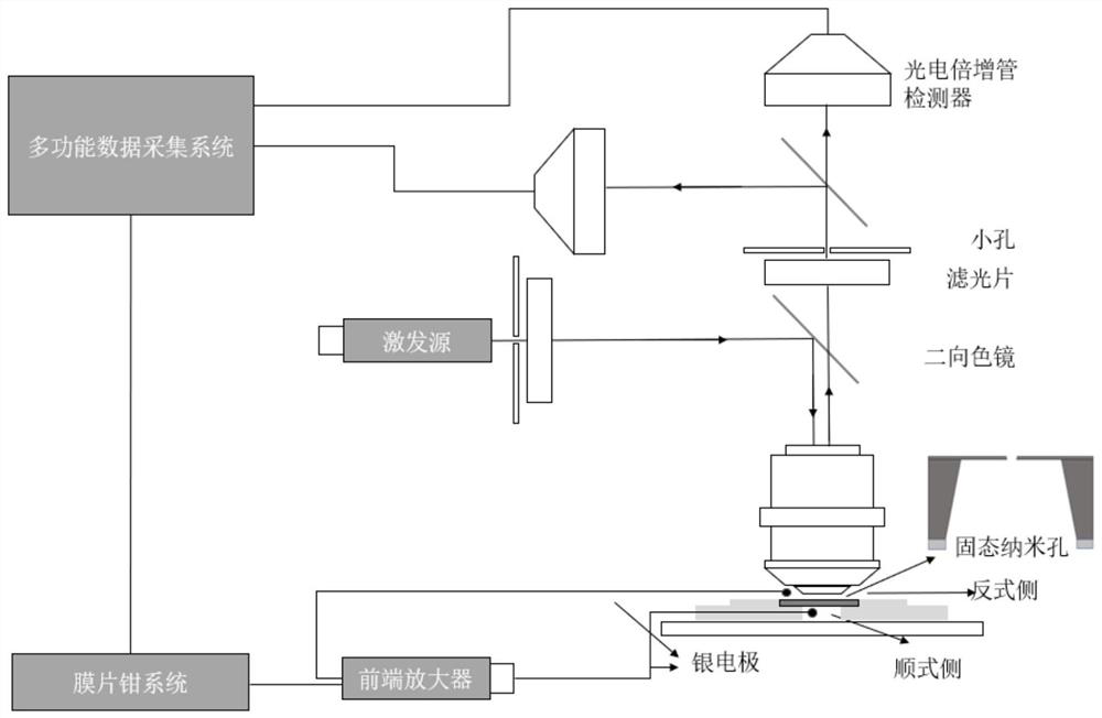
[0022] The preparation of a silicon nitride solid-state nanopore chip, through the process of drilling nanoscale holes on a silicon nitride self-supporting film, comprises the following steps:
[0023] (1) For a silicon wafer coated with silicon dioxide on one side, perform negative photolithography on the silicon dioxide side. After the photolithography on the silicon dioxide side is completed, it is immersed in hydrofluoric acid to remove the silicon dioxide layer and expose the silicon substrate in the middle; then the silicon substrate is etched with tetramethylammonium hydroxide solution under heating conditions to obtain Suspended silicon nitride film.
[0024] (2) Using spherical aberration-corrected transmission electron microscopy, nanopores were prepared on the suspended silicon nitride film. Clamp the above-mentioned silicon nitride self-supporting thin film chip into the sample holder of the transmission electron microscope (place the side with the suspended silic...
Embodiment 2
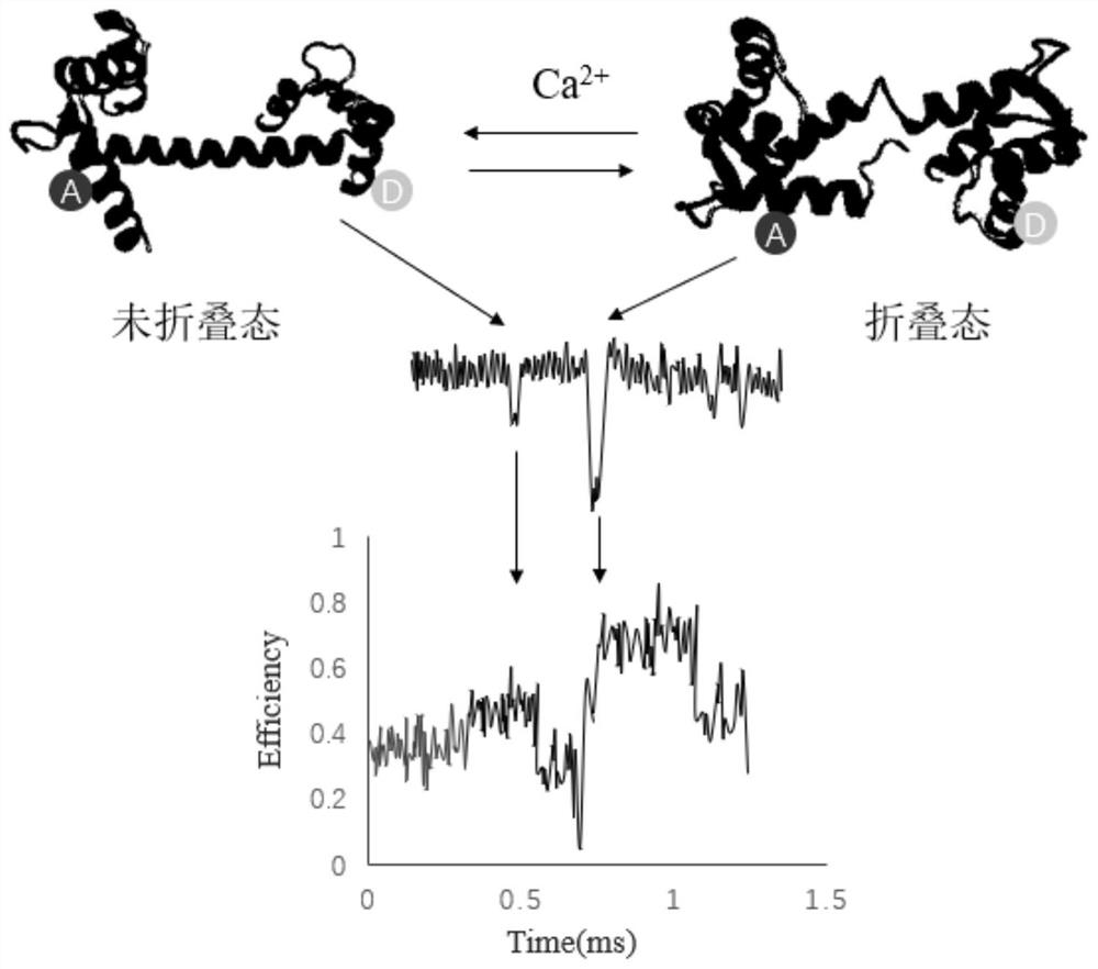
[0028] The platform built according to Example 1 detects different conformational states of calmodulin. Potassium chloride solution was dripped into the cis chamber, and the C-terminal and N-terminal domains of calmodulin were labeled with fluorescent probes Cy3 (fluorescent donor, marked as D) and Cy5 (fluorescent acceptor, marked as A), respectively, And dispersed in potassium chloride solution, drop into the trans side. CaM is a typical protein that can undergo conformational changes after calcium binding. 2+ After binding, CaM undergoes a change in configuration, and its conformation will change from a closed state to an open state, and at the same time expose the hydrophobic region, the surface of the hydrophobic region can bind to the target molecule of regulation, thus playing a vital role in the regulation of metabolic processes role. FRET values changed significantly when calmodulin conformation changed. Applying a positive pressure to the trans side of the solid...
Embodiment 3
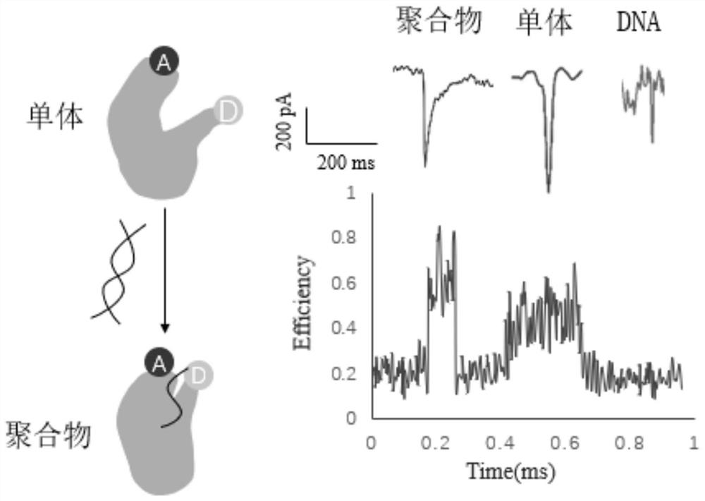
[0030] According to the method of Example 1, the binding of the large Klenow fragment to DNA was detected. The crystal structure shows that when the large Klenow fragment binds to a DNA substrate, the thumb domain undergoes a conformational change, with a change of about 1.2nm. A Klenow large fragment labeled with a Cy3B donor fluorophore (labeled D) and a Cy5 acceptor fluorophore (labeled A) was designed as follows. The donor fluorophore was excited with a 550 nm laser and the fluorescence emission between 560 nm and 750 nm was collected with a time resolution of 50 ms. Imaging buffer contained 50 mm Tris (pH 7.5), 10 mM MgCl2, 100 mM NaCl, 100 μg / ml BSA, 5% glycerol, 1 mM DTT, 1 mM Trolox, 1% glucose oxidase. Corresponding the FRET signal to the nanopore blocking signal in time, the result is as follows image 3 As shown, according to the figure, when the Klenow large fragment forms a polymer with DNA, the conformational change of the thumb domain reduces the distance betw...
PUM
| Property | Measurement | Unit |
|---|---|---|
| wavelength | aaaaa | aaaaa |
Abstract
Description
Claims
Application Information
 - R&D
- Intellectual Property
- Life Sciences
- Materials
- Tech Scout
- Unparalleled Data Quality
- Higher Quality Content
- 60% Fewer Hallucinations
Browse by: Latest US Patents, China's latest patents, Technical Efficacy Thesaurus, Application Domain, Technology Topic, Popular Technical Reports.
© 2025 PatSnap. All rights reserved.Legal|Privacy policy|Modern Slavery Act Transparency Statement|Sitemap|About US| Contact US: help@patsnap.com


