Anti-porcine sapelovirus VP1 protein hybridoma cell strain, monoclonal antibody and application thereof

A hybridoma cell line, monoclonal antibody technology, applied in antiviral immunoglobulin, application, instrument and other directions, can solve the problem of no detection antibody yet

Active Publication Date: 2021-11-05
YANGZHOU UNIV
View PDF4 Cites 1 Cited by
  • Summary
  • Abstract
  • Description
  • Claims
  • Application Information

AI Technical Summary

Problems solved by technology

At present, only prokaryotic expression system is used to express PSV recombina

Method used


Image

  • Anti-porcine sapelovirus VP1 protein hybridoma cell strain, monoclonal antibody and application thereof
  • Anti-porcine sapelovirus VP1 protein hybridoma cell strain, monoclonal antibody and application thereof
  • Anti-porcine sapelovirus VP1 protein hybridoma cell strain, monoclonal antibody and application thereof

Examples


Embodiment 1

[0034] The establishment of embodiment 1 monoclonal antibody hybridoma cell line

[0035] 1 Materials and methods

[0036] 1.1 Experimental materials

[0037] 1.1.1 Main reagents

[0038] HAT and HT selection medium were purchased from Beijing Boaolong Company, HRP goat anti Mouse IgG was purchased from Abcam Life Science Co., Ltd., Freund's complete adjuvant, Freund's incomplete adjuvant, PEG1450 was purchased from Sigma Company, DMEM medium was purchased from Fetal bovine serum was purchased from Gibco Company, Hangzhou Sijiqing Company, goat anti-mouse IgG fluorescent antibody was purchased from Abbkine, Protein A / G immunoprecipitation magnetic beads were purchased from Bimake Company, and fluorescence microscope was purchased from German LeiCa Company.

[0039]1.1.2 Cells, experimental animals, virus strains and serum

[0040] ST-R cells (porcine testicular cells with IFN-β receptor knockout) and SP2 / 0 myeloma cells were cryopreserved in the inventor's laboratory. Fema...

Embodiment example 2

[0071] Implementation Case 2 Characterization of Monoclonal Antibodies

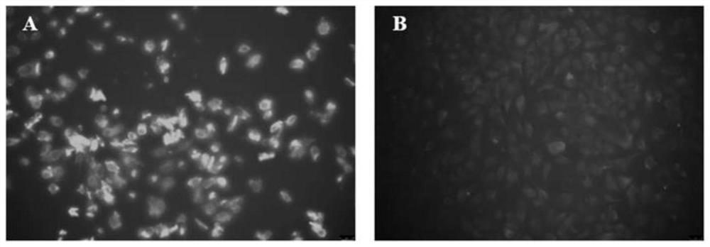
[0072] (1) Indirect immunofluorescence test of monoclonal antibody and PSV

[0073] Spread the ST-R cells in a 24-well plate, inoculate the PSV virus fluid at MOI=0.01, and use uninfected ST-R cells as a negative control. About 16-20 hours after infection, discard the medium, wash with PBS 3 times, add 4% paraformaldehyde to fix at room temperature for 10 minutes, discard the fixative, wash 3 times with PBS, bovine serum albumin (BSA) and Triton-X-1001: 1 Mix and block for 30 minutes, wash with PBS 3 times, add VP1 monoclonal antibody, incubate at 37°C for 1 hour, wash with PBS 3 times, then incubate with Dylight488 goat anti-mouse secondary antibody (1:2000) for 1 hour at 37°C in the dark, wash 3 times with PBS, Observe the results with an inverted fluorescence microscope.

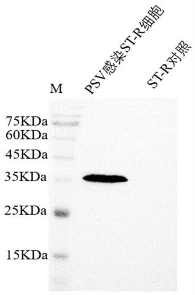
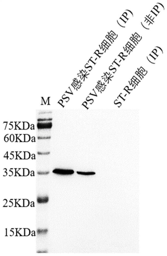
[0074] (2) Western blot to identify the reactivity of monoclonal antibodies

[0075] Inoculate ST-R cells in a 6-well plate, and...

Embodiment example 3

[0078] Implementation Case 3 Monoclonal Antibody Epitope Identification

[0079] (1) Preliminary identification of monoclonal antibody epitopes

[0080] Using the PSV-HuN2 strain as a template, four partially overlapping VP1 protein gene fragments A, B, C, and D were amplified, and each fragment was inserted into the pEGFP-C3 vector through restriction sites, and the positive plasmids constructed were sent to Nanjing Qing Sequencing department. According to the instructions of Entranster-H4000, each recombinant plasmid was transfected into HEK293A cells, and the total protein of the cells was collected after 36 hours. Monoclonal antibody (1:1000) was used as the primary antibody, and HRP Goat Anti-Mouse IgG was used as the secondary antibody. The region of the preliminary antigen epitope is located on the B fragment, and the amino acid position is 30-75aa. As above, segment B was divided into three partially overlapping gene segments B1, B2, and B3, relevant primers were des...


PUM

PropertyMeasurementUnit
Upstream primeraaaaaaaaaa

Abstract

The invention belongs to the technical field of biology, and discloses a hybridoma cell strain secreting a porcine sapelovirus VP1 protein monoclonal antibody. The preservation number of the hybridoma cell strain is CCTCC NO: C2021177, and the hybridoma cell strain is preserved in China Center for Type Culture Collection, Wuhan University, Wuhan, China. The hybridoma cell has stable antibody secretion capacity, the secreted monoclonal antibody has good reaction specificity with the porcine sapelovirus VP1 protein, the antigen epitope recognized by the monoclonal antibody is the 40th-46th amino acid of the porcine sapelovirus VP1 protein, the polypeptide sequence of the antigen epitope is 40PALTAAE46, and the epitope is not reported yet. The hybridoma cell strain lays a good material foundation for researching the etiology and pathogenesis of the porcine sapelovirus in the future.

Description

technical field [0001] The invention belongs to the field of biotechnology, and in particular relates to a hybridoma cell strain expressing a monoclonal antibody to porcine Sapero virus VP1 protein. Background technique [0002] Porcine Sapelovirus (PSV) was first discovered in the UK and was named porcine enterovirus 8 (PEV8) because it was first isolated from the intestinal tract of pigs with diarrhea. PSV is mainly transmitted through the fecal-oral route, and can cause porcine poliomyelitis, diarrhea, pneumonia, reproductive disorders, myocarditis and other multisystem syndromes, and can also manifest as subclinical infections without obvious characteristics. Mixed infection with pestivirus, porcine parvovirus, porcine epidemic diarrhea virus, porcine reproductive and respiratory syndrome virus, and porcine enterovirus has brought serious harm to the pig industry. [0003] Porcine Saperovirus is a microspherical, non-enveloped, single-stranded positive-sense RNA virus b...

Claims


Application Information

Patent Timeline
IPC IPC(8): C12N5/20C07K16/10G01N33/569G01N33/577C12N15/13
CPCC07K16/1009G01N33/56983G01N33/577C07K2317/565C07K2317/56C07K2317/51C07K2317/515C12N2770/32022Y02A50/30
Inventor 陈振海王羽茜牟春晓吴慧光
Owner YANGZHOU UNIV
Why Eureka
Try Eureka