Preparation method of developable high-adhesion drug sustained-release body for nerve block

A nerve block and high-adhesion technology, applied in the biological field, can solve the problem that the position of drug diffusion cannot be effectively displayed, reduce the drug action time, etc., and achieve the effect of improving accuracy and prolonging action time.

Pending Publication Date: 2021-11-12
THE FIRST AFFILIATED HOSPITAL OF ZHENGZHOU UNIV
View PDF4 Cites 0 Cited by
  • Summary
  • Abstract
  • Description
  • Claims
  • Application Information

AI Technical Summary

Problems solved by technology

[0003] However, in this process, although ultrasound can also improve the accuracy of puncture by displaying the position of the puncture needle, it cannot effectively show the position of drug diffusion during and after injection, and the injection of large doses of local anesthetics is always difficult. On the one hand, it will cause uncontrollable nerve damage, on the other hand, the fast drug metabolism rate will also reduce the drug's action time

Method used


Image

  • Preparation method of developable high-adhesion drug sustained-release body for nerve block

Examples


Embodiment 1

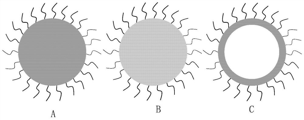
[0045] Preparation of Polydopamine-modified Silk Fibroin with Uniform Spherical Structure for Ultrasonic Imaging Drug Sustained Release

[0046] 1) Preparation of silk fibroin prepolymer solution: silk fibroin is extracted from silkworm cocoons. The freeze-dried silk fibroin was dissolved in hexafluoroisopropanol solvent to prepare silk / hexafluoroisopropanol solution. Then slowly drop required volume of deionized water into 3ml of 15% w / v silk / hexafluoroisopropanol solution, and then mix thoroughly by gently swirling to obtain silk fibroin prepolymer solution.

[0047] 2) Preparation of monodisperse polydopamine-modified silk fibroin microspheres: the preparation of monodisperse silk fibroin microspheres adopts microfluidic technology. The silk fibroin prepolymer solution obtained above is used as the water phase of microfluidics, and the external phase adopts n-hexadecane containing surfactant, collects the generated droplets, and transfers it to a glass bottle, seals and st...

Embodiment 2

[0050] Preparation of the porous structure based on silk fibroin material modified by de-adrenaline polymer, and the preparation of high-adhesive sustained-release drug by ultrasound imaging

[0051] 1) Preparation of silica particles:

[0052] ① Preparation of monodisperse silica nanoparticles: uniformly mix water, tetraethyl orthosilicate, arginine, and cyclohexane to prepare monodisperse silica nanoparticles with a size of 10-20nm; The above particle solution is dispersed in the seed growth reaction system constructed by ethanol and ammonia water, and slowly dripped into the silica prepolymer solution under uniform stirring to obtain monodisperse silica particles of required size.

[0053]②Preparation of silica microsphere template preparation: the monodisperse 200nm silica nanoparticles were centrifuged and purified, and the mass percentage of colloidal nanoparticles in the colloidal nanoparticles solution was adjusted to 20%. The colloidal nanoparticle solution was dispe...

Embodiment 3

[0057] Preparation of Polydopamine-modified Highly Adhesive Drug Sustained-Release Body Based on the Hollow Structure of Polylactic-Glycolic Acid Material by Ultrasound Imaging

[0058] 1) Preparation of polylactic acid-glycolic acid microspheres with hollow structure: Add 0.3g polylactic acid-glycolic acid into 6ml of dichloromethane solvent, mix well, add 4ml of water, stir at 300rpm for 1 hour, then add 50ml of 1 % polyvinyl alcohol solution, stirred at 50rmp for 24 hours to obtain polylactic acid-glycolic acid microspheres with a hollow structure. The polylactic acid-glycolic acid hollow microspheres obtained were centrifuged (3000 rpm, 5 min), rinsed three times with deionized water, and then freeze-dried in a freeze dryer. The obtained powder is further dispersed in water, and further 0.1 g of dopamine and trishydroxymethylaminomethane are added to the microsphere aqueous solution to adjust the pH of the solution to 8.5, and the polydopamine-modified silk fibroin microsp...


PUM


Abstract

The invention discloses an ultrasound developable high-adhesion drug sustained-release body for nerve block. The drug sustained-release body is a micron-order sphere structure of a medical material, the diameter of the sphere is 1-50 [mu] m, the sphere adhesion material realizes in-vivo adhesion through surface modification of adhesion molecules, and under the ultrasonic guidance, the development under the ultrasonic is realized by utilizing the scattering effect of the device on mechanical waves. The ultrasonic developable drug sustained-release body can accurately locate the action position of the drug, the action time of the drug can be prolonged through the adhesion and sustained-release effects of the drug sustained-release body, and the accuracy of nerve block treatment and the bioavailability of the drug are improved.

Description

technical field [0001] The invention relates to the field of biotechnology, in particular to a high-adhesive drug slow-release body that can be developed for nerve block and a preparation method thereof. Background technique [0002] Nerve blocks are a major means of effective treatment of a variety of acute and chronic pain. Ultrasound-guided nerve block uses real-time ultrasound to clearly display most of the peripheral nerves and their surrounding anatomical structures, so as to precisely position and inject anesthetic drugs to the treatment site, and has achieved good results in pain treatment. [0003] However, in this process, although ultrasound can also improve the accuracy of puncture by displaying the position of the puncture needle, it cannot effectively show the position of drug diffusion during and after injection, and the injection of large doses of local anesthetics is always difficult. On the one hand, it will cause uncontrollable nerve damage, on the other ...

Claims


Application Information

Patent Timeline
Patent Type & Authority Applications(China)
IPC IPC(8): A61K49/22
CPCA61K49/223A61K49/221
Inventor 杨建军刘盼苗尚会杰叶皓天
Owner THE FIRST AFFILIATED HOSPITAL OF ZHENGZHOU UNIV
Why Eureka
Try Eureka