Modeling method of mouse hypertensive cerebral hemorrhage model

A hypertensive cerebral hemorrhage and mouse technology, applied in the field of biomedicine, can solve problems such as the inability to simulate the pathological changes and disease outcomes of cerebral hemorrhage, the inability to form a hematoma, and the difficulty of puncturing.

Pending Publication Date: 2021-11-16
XUZHOU MEDICAL UNIV
View PDF0 Cites 1 Cited by
  • Summary
  • Abstract
  • Description
  • Claims
  • Application Information

AI Technical Summary

Problems solved by technology

However, the volume of hematoma is not easy to control, showing diffuse hemorrhage, which is not conducive to experimental controlled research
And the high mortality rate limits the application of this model
In addition, collagenase itself has neurotoxicity, and will amplify the inflammatory response, which affects the establishment of the model, and cannot well simulate the pathological changes and disease outcome of human cerebral hemorrhage.
Another intracranial autologous blood injection model of cerebral hemorrhage, this method is to draw autologous blood from rats and inject it into the basal ganglia area using a brain locator. It has been reported in the literature that blood is drawn from the tail vein and periorbital of rats, but the blood drawn is venous blood, and arterial blood forms a hematoma in hypertensive cerebral hemorrhage, so this method cannot simulate clinical hypertensive cerebral hemorrhage well.
There is also the method of tail-cutting to obtain blood, which is to cut off the tail end of the mouse to obtain blood, but this method obtains mixed arterial and venous blood, not pure arterial blood.
In addition, there are reports in the literature that blood can be obtained by puncturing the tail artery of rats. This method is suitable for rats, but the tail artery of mice is located in the deep part of the ventral surface, which is very thin and difficult to puncture. Meet the experimental needs
[0004] To sum up, the existing modeling technology has the following shortcomings and deficiencies: (1) It is impossible to find the best puncture site and puncture channel in the mouse body, so as to extract a sufficient amount of arterial blood from the mouse body for the experimental study. need
(2) The survival rate of mice cannot be guaranteed after puncture and blood collection
(3) A stable hematoma cannot be formed, which cannot provide guarantee for subsequent experiments, is not conducive to experimental control studies, and cannot well simulate the pathological changes and disease outcomes of human cerebral hemorrhage

Method used


Image

  • Modeling method of mouse hypertensive cerebral hemorrhage model
  • Modeling method of mouse hypertensive cerebral hemorrhage model
  • Modeling method of mouse hypertensive cerebral hemorrhage model

Examples


Embodiment 1

[0040] A method for building a mouse hypertensive cerebral hemorrhage model, the specific steps are as follows:

[0041] (1) Blood collection by left ventricular puncture: the mice were anesthetized by isoflurane gas induction, the mice were placed in the supine position on the cardboard, the limbs were fixed with medical adhesive tape, the cotton thread was buckled over the front teeth of the mice, and pulled Fix the mouse so that the chest and upper abdomen of the mouse are fully exposed. Use a cotton swab dipped in 75% standard medical alcohol to disinfect the chest and upper abdomen on a large scale, touch the strongest heartbeat (apex) of the chest with the left hand, and then use a 1mL syringe at the angle between the xiphoid process of the sternum and the ribs at an angle of 10° Puncture at the point with the strongest heartbeat at ~20°, draw blood while withdrawing the needle, and quickly transfer the arterial blood to a 1.5mL EP tube for later use.

[0042] (2) Expos...

Embodiment 2

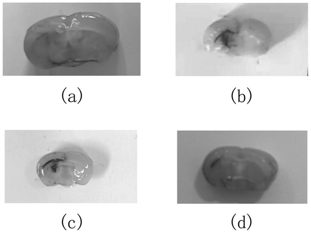
[0048] Example 2 Coronal view of hematoma after hypertensive cerebral hemorrhage modeling in mice

[0049] Ten 5- to 7-week-old male Kunming mice (derived from swiss mice), weighing 20 to 30 g, were divided into two groups (n=5), respectively Sham and ICH groups, raised in a quiet environment and given free access to water Feed. The sham group did not draw autologous blood, but only inserted the microsampler 3.7 mm from the drill hole to the ventral side, kept the needle in place for 10 minutes before pulling it out, then pulled it back at a speed of 1 mm / min, and sealed it with bone wax Holes were drilled and the skin was sutured. The ICH group was performed according to the specific steps of the modeling method described in Example 1. After modeling, the coronal plane of Sham group and the coronal plane of hematoma in ICH group were observed. Coronal incision was made on the coronal plane of ICH group with the injection point as the center point, and 1, 2, and 3 days were ...

Embodiment 3

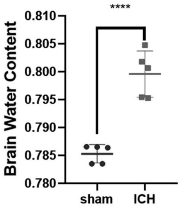
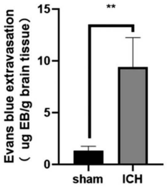
[0051] Example 3 Determination of degree of brain edema after hypertensive intracerebral hemorrhage modeling in mice

[0052] Ten 5- to 7-week-old male Kunming mice, weighing 20-30 g, were divided into two groups (n=5), namely the Sham group and the ICH group, and were reared in a quiet environment with free access to water and food. The sham group did not draw autologous blood, but only inserted the microsampler 3.7 mm from the drill hole to the ventral side, kept the needle in place for 10 minutes before pulling it out, then pulled it back at a speed of 1 mm / min, and sealed it with bone wax Holes were drilled and the skin was sutured. The ICH group was performed according to the specific steps of the modeling method described in Example 1. Then, using the dry-wet weight method, the mice in each group were injected intraperitoneally with 10% chloral hydrate for 24 hours to induce anesthesia, the brain was decapitated, the olfactory bulb, cerebellum and brainstem were removed...


PUM


Abstract

The invention discloses a modeling method of a mouse hypertensive cerebral hemorrhage model, which comprises the following steps of: puncturing and taking blood from the left ventricle of a mouse, exposing a skull surgery field by wiping and cleaning soft tissues covering the skull, drilling after determining a needle insertion point on the surface of the skull, injecting autologous blood at the drilling position through a brain stereotaxic instrument, and then injecting the autologous blood into the mouse hypertensive cerebral hemorrhage model; and after injection is completed, sealing the drilled hole with bone wax, disinfecting the incision, and suturing the skin. According to the method, arterial blood is taken through left ventricle puncture, the survival rate of a mouse is not reduced on the premise that sufficient arterial blood is extracted, autologous arterial blood is accurately injected back into the tail shell core of the basal segment in the brain through a mouse brain locator, and stable hematoma is formed. The method provides guarantee for subsequent experiments, is beneficial to experimental contrast research, is very similar to clinical pathological changes and disease outcome of hypertension patients, and has important significance on disease outcome and treatment schemes after cerebral hemorrhage.

Description

technical field [0001] The invention belongs to the technical field of biomedicine, and in particular relates to a modeling method of a mouse hypertensive cerebral hemorrhage model. Background technique [0002] Intraparenchymal hemorrhage is the second most common type of stroke, with the characteristics of high morbidity, high mortality and high disability rate, accounting for about 20% to 30% of all strokes in my country. Hypertensive intracerebral hemorrhage (intracerebral hemorrhage, ICH) often occurs in the putamen and internal capsule of the basal ganglia, accounting for about 70% of ICHs. Putamen hemorrhage often invades the internal capsule, often causing contralateral hemiplegia and hemisensory loss and homotropic hemianopsia. Cerebral hemorrhage can cause cerebral edema, damage to the integrity of the blood-brain barrier, limb sensorimotor impairment, and neurological deficits. Therefore, it is of great significance to study the disease outcome and treatment pla...

Claims


Application Information

Patent Timeline
Patent Type & Authority Applications(China)
IPC IPC(8): A61D1/00
CPCA61D1/00
Inventor 胡泊荣良群张清秀
Owner XUZHOU MEDICAL UNIV
Why Eureka
Try Eureka