
Target positioning and quantitative detection method based on DNA spherical nanostructure imaging

A quantitative detection method and nanostructure technology, applied in the determination/inspection of microorganisms, biochemical equipment and methods, and labels used in chemical analysis, etc., can solve the problem of limited, ineffective detection of low-abundance and rare targets, and spontaneous detection of samples. Issues such as fluorescence and light scattering

Pending Publication Date: 2021-11-23
CHINA PHARM UNIV
View PDF0 Cites 0 Cited by
  • Summary
  • Abstract
  • Description
  • Claims
  • Application Information

AI Technical Summary

Problems solved by technology

In addition, immunofluorescence methods have no signal amplification or limited signal amplification based on secondary antibody methods, so their detection sensitivity is not high enough to effectively detect low-abundance rare targets
In addition, when performing fluorescence detection on biological samples, there will be problems with autofluorescence and light scattering in the sample, which further limits the use of immunofluorescence methods using fluorescence intensity quantification for the characterization of low-abundance targets.

Method used


Image

  • Target positioning and quantitative detection method based on DNA spherical nanostructure imaging
  • Target positioning and quantitative detection method based on DNA spherical nanostructure imaging
  • Target positioning and quantitative detection method based on DNA spherical nanostructure imaging

Examples


Embodiment 1

[0057] (1) Experimental materials and reagents:

[0058] MDA-MB-231 cell line was purchased from Shanghai ATCC cell bank; cell culture grade phosphate buffered saline solution (PBS solution, without calcium chloride, magnesium chloride, 1×PBS pH 7.4) was purchased from the United States DMEM medium (containing penicillin-streptomycin double antibody) was purchased from Keygen; trypsin (Trypsin) was purchased from the United States Sterile fetal bovine serum (FBS) was purchased from Natocor-Industria, Argentina Bovine serum albumin (bovine serum albumin, BSA) was purchased from American Amresco Company; Glass Bottom Cell Culture Dish cell culture dish was purchased from Wuxi Nice Biotechnology Co., Ltd.; biotin, formamide, dimethyl sulfoxide (DMSO) And polyethylene glycol tert-octyl phenyl ether (Triton X-100) was purchased from Sigma-Aldrich Company of the United States; 4′, 6-diamidino-2-phenylindole (DAPI) was purchased from Wuhan Boster (Boster ) company; all oligonucl...

Embodiment 2

[0070] Example 2 Oligonucleotide labeling and corresponding design of single-stranded DNA molecules used to form circular templates

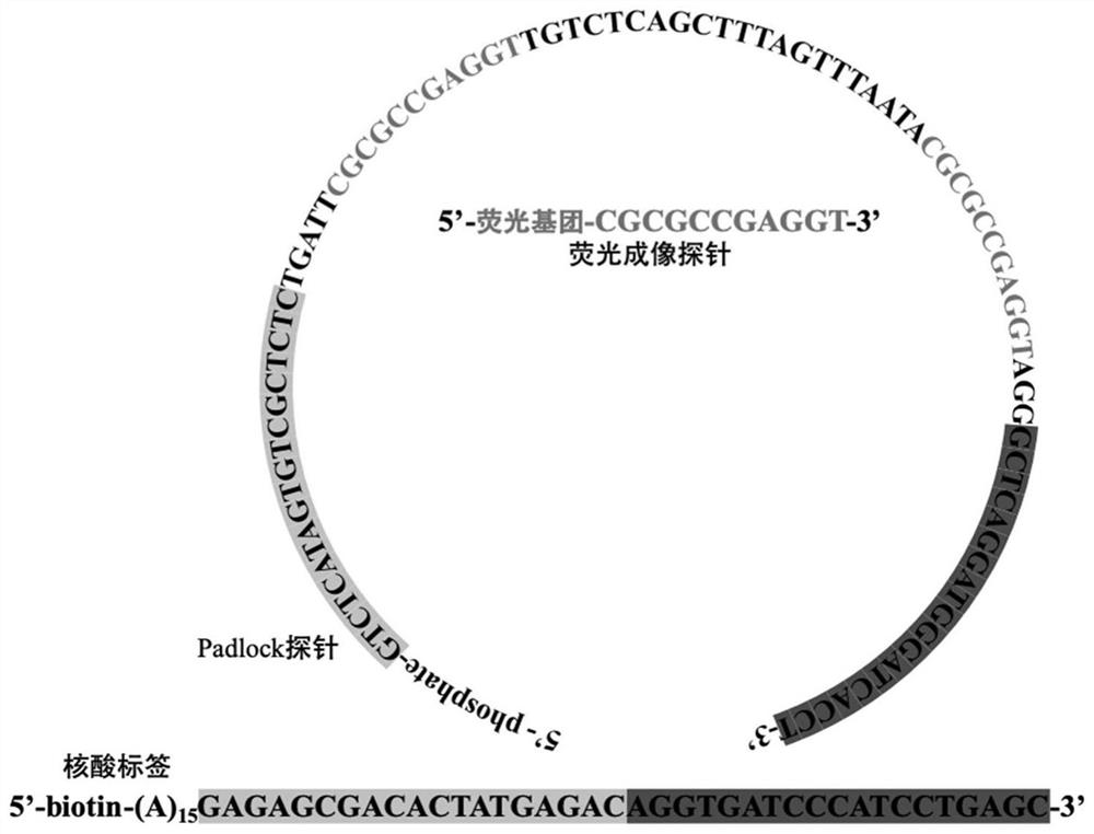
[0071] see image 3 , in this example, see Example 1 for specific experimental materials and reagents, the 5' end of the oligonucleotide tag 1 is biotinylated, so that the tag and the target-specific binding molecule are passed through biotin- Streptavidin system for bridging. The single-stranded DNA molecule (Padlock molecule 1) contains a complementary part to the nucleic acid tag to form a circular template, and also contains a template sequence for the generation of the hybridization sequence of the fluorescently labeled probe 1, so that only when the oligonucleotide tag 1 is amplified In order to generate a complementary hybridization sequence with the fluorescent label probe 1, the specificity of the fluorescent signal is ensured.

Embodiment 3

[0072] Example 3 Glyceraldehyde-3-phosphate dehydrogenase (glyceraldehyde-3-phosphate dehydrogenase) protein detection

[0073] see Figure 4 , in this embodiment, specific experimental materials and reagents, cell culture experimental steps, contents and conditions, oligonucleotide tag-labeled antibody modification experimental steps, contents and conditions refer to Example 1, will be fixed with 4% paraformaldehyde The final MDA-MB-231 cell sample is in contact with the target-specific binding coupler, so that it can be stably and specifically bound to the sample, and the target is formed under the action of rolling circle replication mediated by the nucleic acid label on the target-specific binding coupler. Specific DNA nanospheres can be hybridized with specific fluorescently labeled probes to form hybrids, forming fluorescent bright spots that are clearly distinguished from the background under a fluorescent microscope.

[0074]Target detection experimental steps, conten...


PUM


Abstract

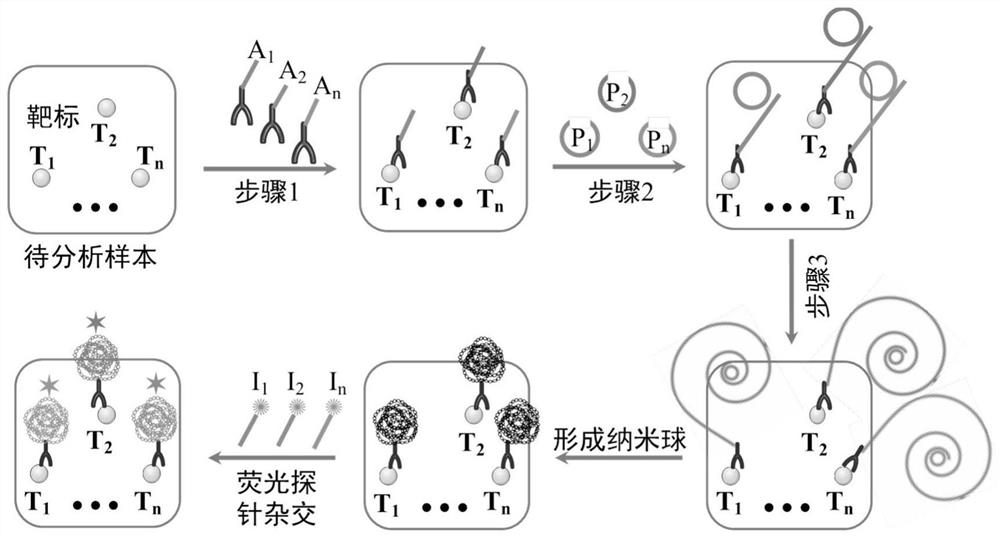
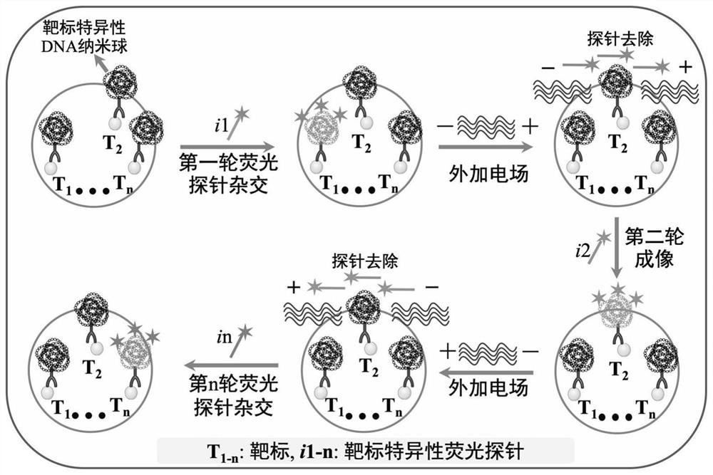
The invention discloses a target positioning and quantitative detection method based on DNA spherical nanostructure imaging. The target quantitative detection method relates to signal amplification detection, in particular to high-sensitivity detection of a low-abundance target in a sample; the multiple detection and multiple numbers of the target break through the limitation that a common fluorescence microscope can only detect 3-5 targets at the same time. The key content of the method relates to a case that a target specific detection object, such as an antibody, is connected with a single-chain oligonucleotide tag, then the single-chain oligonucleotide tag is amplified by using DNA loop-mediated rolling circle amplification, and the amplified nucleic acid can spontaneously form a spherical DNA nanostructure under the action of intramolecular hybridization; by designing a ring forming probe sequence, the spherical DNA nanostructure formed by a rolling circle amplification product can be hybridized with a fluorescently-labeled specific nucleic acid probe, so that the spherical DNA nanostructure shows a bright spot under a fluorescence microscope; and by positioning and counting the bright spot, positioning and digital quantitative detection of a to-be-detected target are implemented.

Description

technical field [0001] The invention belongs to the field of positioning and quantitative detection, in particular to a multi-target positioning and quantitative detection method in cells and tissues based on DNA spherical nanostructure imaging. Background technique [0002] Proteins and other biomolecules are important components of all cells and tissues in the human body, and are important participants in the execution of human physiological functions. More and more studies have shown that the occurrence and development of diseases are closely related to the interaction of some proteins and other biomolecules in tissue cells. Therefore, it is very important to elucidate the types and content changes of proteins and other biomolecules in samples such as tissue cells to deeply understand the occurrence and development of diseases. [0003] It is worth noting that biomolecules such as proteins synthesized by cells need to be transported to specific subcellular locations to pl...

Claims


Application Information

Patent Timeline
IPC IPC(8): C12Q1/682C12Q1/6804G01N33/53G01N33/573G01N33/68
CPCC12Q1/682C12Q1/6804G01N33/53G01N33/5308G01N33/573G01N33/68G01N2458/10G01N2333/90203G01N2333/70525C12Q2521/501C12Q2531/125C12Q2525/151C12Q2563/107C12Q2565/601
Inventor 邹秉杰王琛宋沁馨
Owner CHINA PHARM UNIV
Learn More