Method for E, coli to express lysostaphin in high efficiency via external secretion

一种溶葡萄球菌酶、大肠杆菌的技术,应用在大肠杆菌高效外分泌表达溶葡萄球菌酶领域,能够解决回收率高、不易清除、影响产品质量等问题,达到纯化简单、回收率高、污染少的效果

Active Publication Date: 2007-01-24
SHANGHAI HI TECH UNITED BIO TECHCAL RES
View PDF3 Cites 8 Cited by
  • Summary
  • Abstract
  • Description
  • Claims
  • Application Information

AI Technical Summary

Problems solved by technology

In this invention, lysostaphin is expressed in the form of inclusion bodies in Escherichia coli, and the expressed lysostaphin is stored in the cells in the form of insoluble inclusion bodies, and the cells must be broken before they can be separated. During the separation process, a protein denaturant is used, and finally the protein must be refolded. During the refolding process, many molecules will be misconnected to form molecules with abnormal structures. At the same time, the residual protein of the bacteria is difficult to remove during the production of genetic engineering drugs. Substances that are likely to affect product quality
[0006] At present, foreign countries have not been able to achieve efficient exocrine expression of lysostaphin gene in Escherichia coli. The advantage of exocrine expression is that the expression product exists in the medium in the final active form, without steps such as renaturation of inclusion bodies; Purification is relatively simple, the recovery rate is high; host bacterial protein contamination is less

Method used


Image

  • Method for E, coli to express lysostaphin in high efficiency via external secretion
  • Method for E, coli to express lysostaphin in high efficiency via external secretion
  • Method for E, coli to express lysostaphin in high efficiency via external secretion

Examples


Embodiment 1

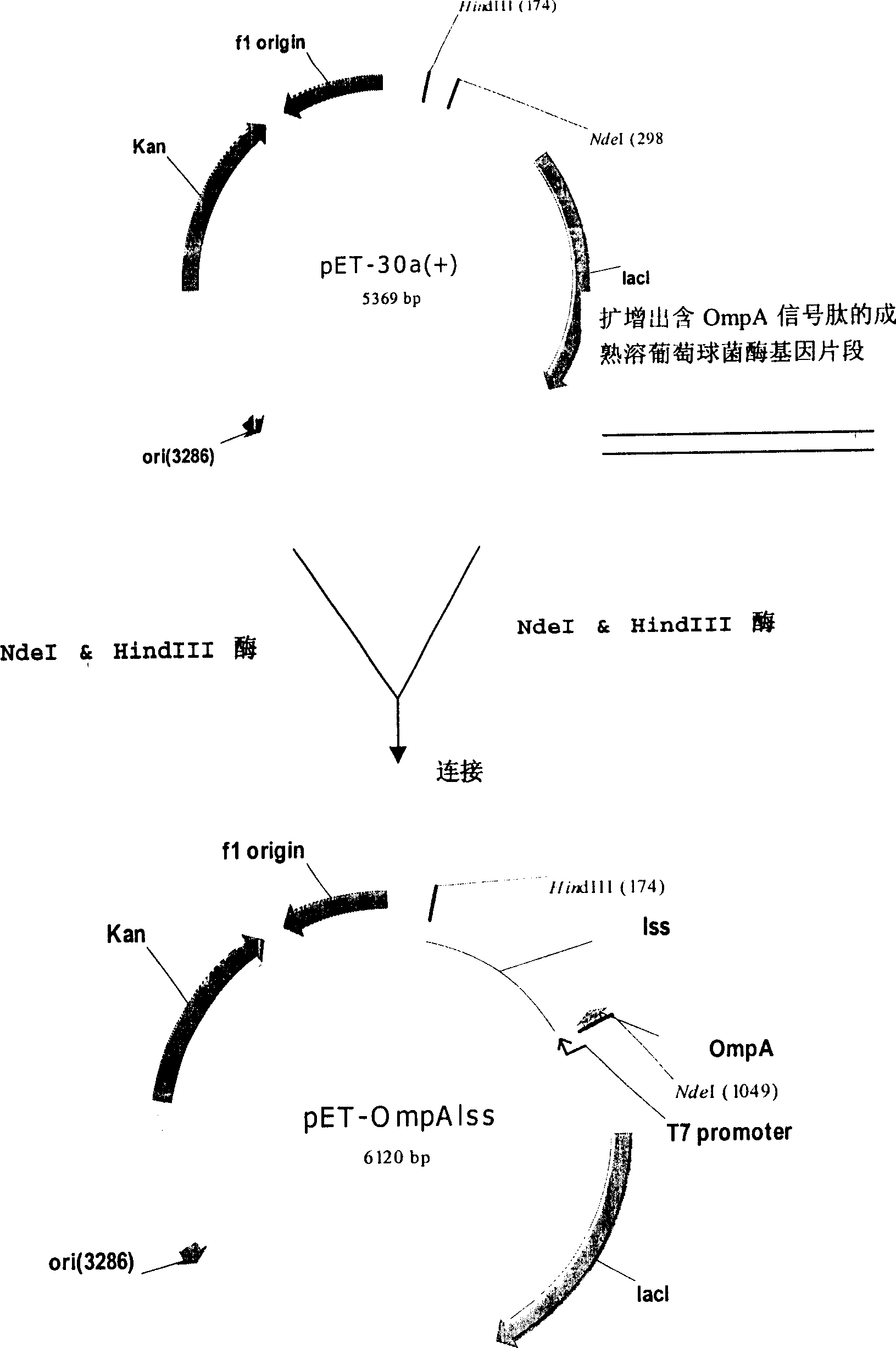
[0044] Example 1: Construction of recombinant lysostaphin secretion expression vector, namely OmpA signal peptide and mature lysate

[0045] Cloning of staphylococcal enzyme fusion gene.

[0046] First design and synthesize the following oligonucleotide sequence:

[0047] SEQ.ID.NO:1 5'-

[0048] TACAT ATGAAAAAGACAGCTATCGCGATTGCAGTGGCACTGGCAGGTTTCG

[0049] CTACCGTCGCTCAGGCT GCTGCAACACATGAACATTCAGCAC-3′

[0050] SEQ.ID.NO: 2 5'-CCAAAGCTTCAACTTTAGGAATGAG-3'

[0051] In the designed SEQ.ID.NO:1, the gene sequence of the NdeI restriction endonuclease site (italicized part) and the gene sequence of the OmpA signal peptide (underlined part) are added to the 5'end;

[0052] In the designed SEQ.ID.NO: 2, the gene sequence of the HindIII restriction endonuclease site (in italics) is added to the 5'end.

[0053] Using the total DNA of Staphylococus Simulans (NRRL B-2628) as the template, SEQ.ID.NO:1 and SEQ.ID.NO:2 as a pair of primers, using pyrobest enzyme (a high-fide...

Embodiment 2

[0064] Example 2: Establishment of recombinant lysostaphin secretory expression engineering bacteria

[0065] The recombinant plasmid pET-OmpAlss was transformed into E. coli TOP10, JM109 (DE3), BL21 (DE3), respectively. Pick positive clones, insert 50ml LB liquid medium (containing 30mg / ml kanamycin) and shake culture at 37℃, cultivate until the optical density value (wavelength 600nm) is 0.6, then add IPTG to the final concentration of 0.05mM induced protein Expression, continue to culture for 3 hours, centrifuge the fermentation broth, and determine the lysostaphin activity of the supernatant by colorimetry.

Embodiment 3

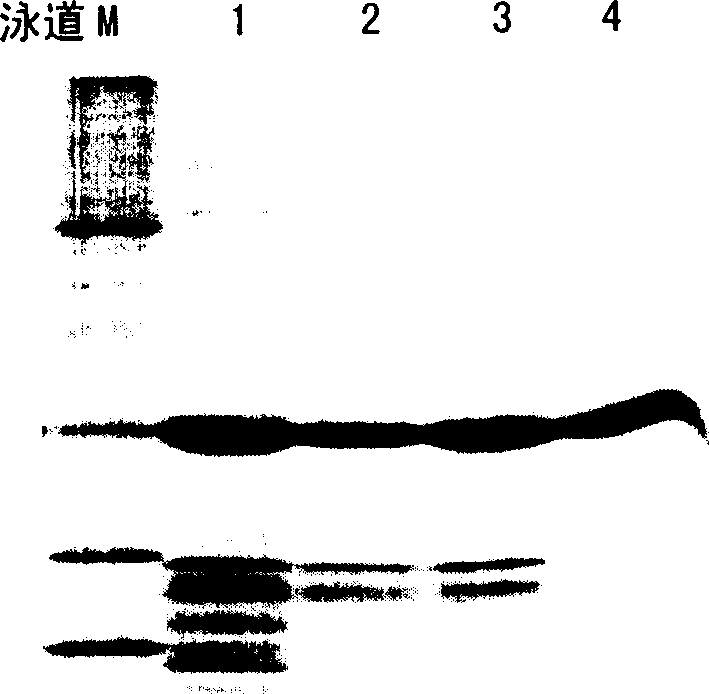
[0066] Example 3 Determination of Lysostaphin Activity by Colorimetry

[0067] The lysostaphin expression activity of TOP10, JM109 (DE3), BL21 (DE3) containing the recombinant plasmid pET-OmpAlss in Example 2 was determined by colorimetry. The specific determination method is as follows:

[0068] 1 principle

[0069] The quantitative detection of lysostaphin enzyme is colorimetric method, using Staphylococcus aureus cell wall peptidoglycan (KNR-PG) coupled with KNR brilliant blue dye as the color source substrate, according to the quantitative release of the enzyme during the action of the enzyme After removing the unreacted insoluble substrate of the small molecule soluble fragment product of the KNR dye group, the supernatant is colorimetrically determined to determine the enzyme activity. This method is simple and easy to implement, sensitive and intuitive.

[0070] 2 Instruments and reagents

[0071] 2.1 Instrument:

[0072] 2.1.1 Ultraviolet-visible spectrophotomet...


PUM


Abstract

The present invention provides method for E. coli to express lysostaphin in high effect via external secretion. The method includes cloning coding signal peptide suitable for secreting expression in E. coli in front of the gene sequence of partially or completely mature lysostaphin and connecting to promoter to constitute the expression vector; transforming the expression vector to E. coli and culturing fermentation; and separating lysostaphin from the supernatant of the fermented liquid. The external secretion expression has the advantages of the expression product existing in the culture medium in ultimate active form and without need of inclusion body renaturation and other steps; simple purification from culture medium supernatant and high recovering rate; and less host bacteria protein pollution.

Description

Technical field [0001] The present invention relates to the field of using recombinant DNA technology to produce protein or polypeptide drugs. More specifically, the present invention relates to a method for producing lysostaphin through exocrine expression of Escherichia coli. Background technique [0002] Lysostaphin was first discovered in the culture of Staphylococcus simulans by Schindler and Schuhard in 1964 (US Patent, Patent No. 3,278,378; 1966), with a molecular weight of 27kDa and containing 246 amino acids. Its mechanism of action is to lyse the glycine pentapeptide bond bridge in the peptidoglycan of the Staphylococcal cell wall, thereby destroying the integrity of the cell wall and dissolving the bacteria. It is an endopeptidase. In the 1980s, the lysostaphin gene was expressed in bacteria such as Escherichia coli, Bacillus subtilis and Bacillus sphaericus through molecular cloning methods at home and abroad. Due to its unique bactericidal mechanism, it can quickly l...

Claims


Application Information

Patent Timeline
Patent Type & Authority Applications(China)
IPC IPC(8): C12N15/70C12N9/36C12N1/21
CPCC07K2319/02C12N9/52Y02A50/30C12N15/00C12N15/09C12N1/20C12P21/02
Inventor 黄青山陆海荣陆婉英
Owner SHANGHAI HI TECH UNITED BIO TECHCAL RES
Why Eureka
Try Eureka