Histoengineering bone and its making process

A technology for tissue engineering bone and cancellous bone, which is applied in the field of biomedical tissue engineering and can solve problems such as the inability to achieve large-scale preparation and marketing

Active Publication Date: 2007-06-06
THE FIRST AFFILIATED HOSPITAL OF THIRD MILITARY MEDICAL UNIVERSITY OF PLA
View PDF0 Cites 23 Cited by
  • Summary
  • Abstract
  • Description
  • Claims
  • Application Information

AI Technical Summary

Problems solved by technology

[0005] However, the current tissue-engineered bones use the patient's own cells as seed cells. The autologous seed cells determine the individualized preparation mode of these tissue-engineered bones. It is required that each application unit must be equipped with a dedicated purification site, professional equipment and personnel. Need to wait for more than 20 days for the proliferation of autologous seed cells and the preparation of tissue engineering products, which cannot achieve large-scale preparation and marketing

Method used


Image

  • Histoengineering bone and its making process
  • Histoengineering bone and its making process
  • Histoengineering bone and its making process

Examples


Embodiment 1

[0035] Example 1 Preparation process of tissue engineered bone constructed by allogeneic MSCs

[0036] 1. Obtain allogeneic red bone marrow: obtain red bone marrow by bone marrow puncture from a healthy donor, and anticoagulate with an equal amount of 1.0 U / ml heparin saline.

[0037] 2. Isolation and cultivation of primary bone marrow mesenchymal stem cells (MSCs): add Percoll separation medium with a density of 1.073g / mL to the diluted bone marrow sample, centrifuge at 400g for 20min, absorb the milky white cloudy nucleated cell layer in the middle, and add D-Hanks Liquid 20ml Gently blow and rinse with 2×10 5 Inoculate the cells at a cell density of / cm2 into plastic culture bottles, add 5ml of complete medium containing 15% fetal bovine serum to each bottle, and incubate in a 37°C, 5% CO2, saturated humidity incubator. Change the medium after 48 hours, and change the medium every other day thereafter. After most of the cell colonies are confluent (usually 10-12 days), su...

Embodiment 2

[0044]Example 2 Preparation process of tissue engineered bone constructed by allogeneic osteoblasts

[0045] 1. Obtain allogeneic periosteum: take out fresh periosteum tissue from a healthy donor through aseptic surgery.

[0046] 2. Isolation and culture of primary allogeneic osteoblasts: Cut the fresh periosteum into small pieces with a side length of about 1mm, digest with 0.1% collagenase for about 40 minutes at 37°C, and stop the reaction with an equal amount of serum-containing medium. After repeated pipetting, inoculate into plastic culture bottles at a cell density of 2×10⁵ / cm2, add 5ml of complete medium containing 15% fetal bovine serum to each bottle, and incubate in an incubator at 37°C, 5% CO2, and saturated humidity. Change the medium after 48 hours, and change the medium every other day thereafter. After most of the cell colonies are confluent (usually 8-10 days), subculture is carried out immediately. Cells of passage 3-5 were collected for later use.

[0047...

Embodiment 3

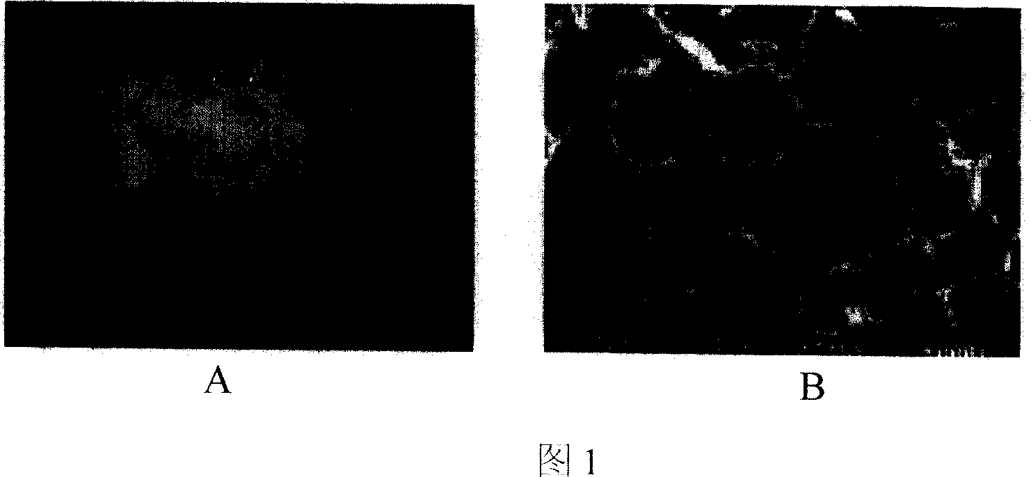
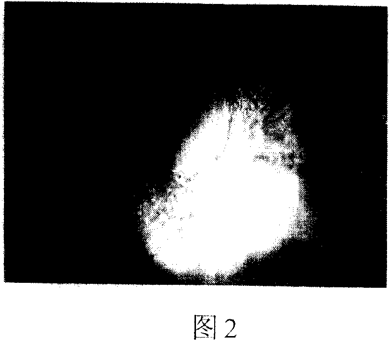
[0051] Example 3 In vitro detection of the above-mentioned demineralized bone matrix (DBM) raw materials:

[0052] Referring to accompanying drawings 1 and 2, the decalcified bone matrix tissue engineering scaffold material is irregular porous solid particles, white or light yellow, without visible impurities, and a particle size of about 8 mm; the rest of the scaffold materials, such as tricalcium phosphate (TCP ), coral, and polylactic acid (PLA) all conform to the three-dimensional shape and external dimensions of their respective specifications. Histological observation: The cancellous bone part of each material is observed under a microscope as a mesh-like structure, with clean pores, smooth walls, and no obvious content. The pore size is 20-300um.


PUM

PropertyMeasurementUnit
particle diameteraaaaaaaaaa

Abstract

The present invention relates to histoengineering bone comprising porous histoengineering bone rack material, composite heterogene seed cell and / or bioactive factor. The histoengineering bone is constituted through treating heterogene cancellous bone via hypotonic solution and ultrasonic cleaning, complete or partial decalcification, defatting, antigen eliminating, etc to obtain high porosity rack material; compounding mesenchyme stem cell, osteoblast and other seed cell and / or bone morphogonetic protein of the heterogene, vascular endothelial growth factor, antibacterial medicine and other bioactive factors; and applying human serum or no serum culture medium following culturing in ox serum culture medium to reduce heterologous serum residue so as to constitute histoengineering bone product ultimately. The histoengineering bone has high performance and is used as bone repairing material clinically.

Description

technical field [0001] The invention belongs to the technical field of biomedical tissue engineering, and in particular relates to a tissue engineering bone used as a clinical bone repair material and a manufacturing method thereof. technical background [0002] Various bone defects caused by severe trauma, tumor resection, bone and joint tuberculosis and infection, congenital bone and joint diseases, and osteoarthritis are very common. The demand is also huge. [0003] Autologous bone transplantation is currently a more effective method for the clinical treatment of bone defects, but the source is limited and will cause new bone defects in the donor site, which will cause additional physical and psychological trauma to the patient, and is a traumatic repair model; The time required for crawling replacement in the body is long, and the incidence of bone nonunion is high; although artificial substitute materials such as metals, ceramics or polymer materials are also used in ...

Claims


Application Information

Patent Timeline
Patent Type & Authority Applications(China)
IPC IPC(8): A61L27/54A61L27/38
Inventor 许建中周强王序全何清义罗飞
Owner THE FIRST AFFILIATED HOSPITAL OF THIRD MILITARY MEDICAL UNIVERSITY OF PLA
Why Eureka
Try Eureka