Device and method or three-dimensional spatial localization and functional interconnection of different types of cells
a technology which is applied in the field of three-dimensional spatial localization and functional interconnection of different types of cells, and can solve the problems of difficult to simulate their cumulative function and anatomically distan
- Summary
- Abstract
- Description
- Claims
- Application Information
AI Technical Summary
Benefits of technology
Problems solved by technology
Method used
Image
Examples
example 2
Polydimethylsiloxane Based Device
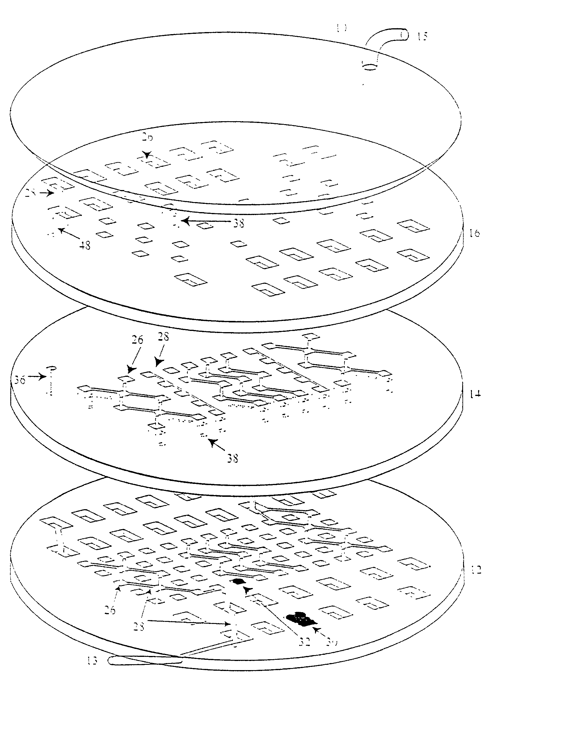
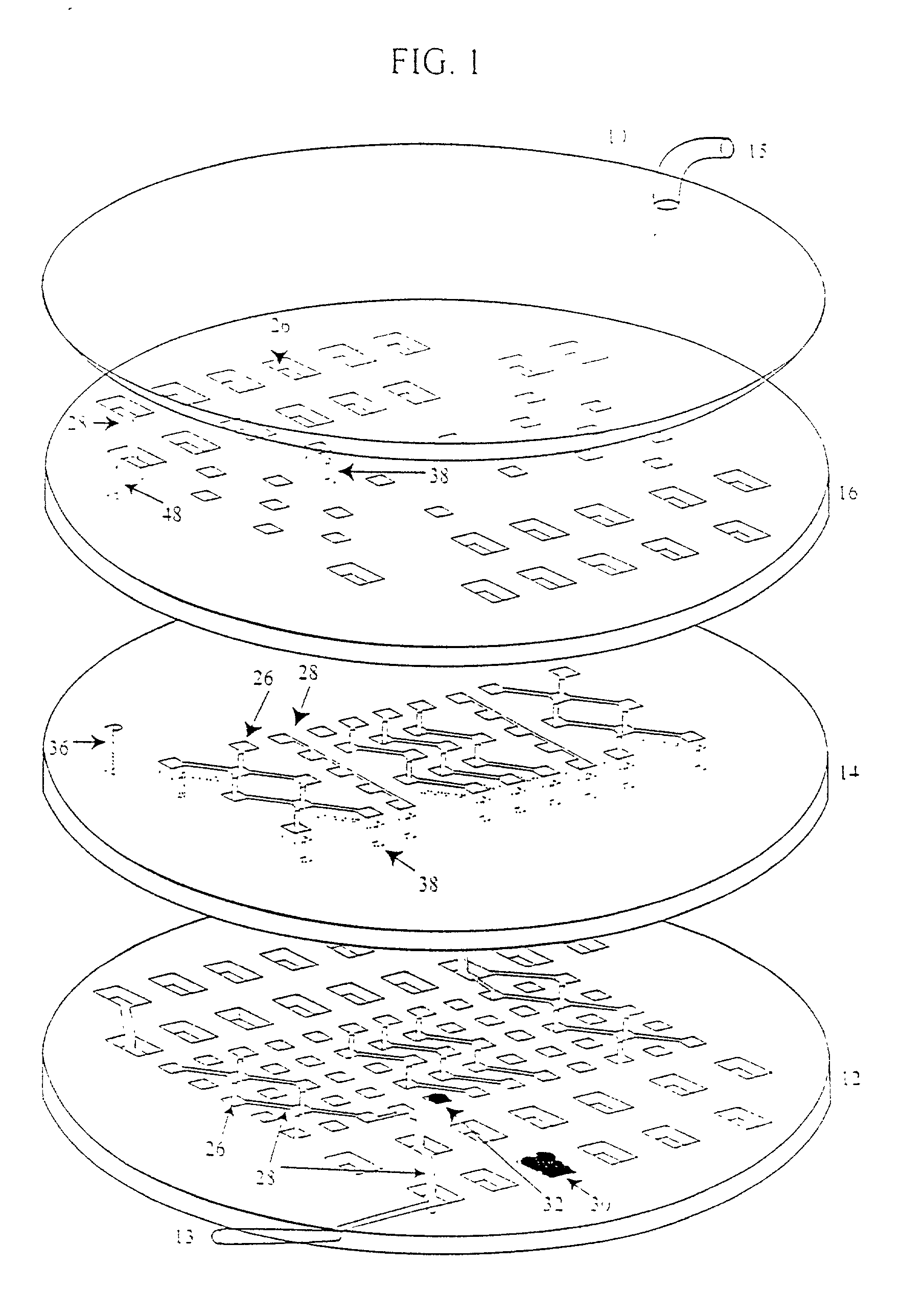
[0247] This example describes fabrication of a polydimethylsiloxane (PDMS) variant of the three-dimensional device.
[0248] Templates for cell isolation arrays are constructed using standard photolithographic methods as described in Example 1.
[0249] Silicon wafers are cleaned of residual organic material in a piranha solution (9:1, H.sub.2SO.sub.4:H.sub.2O.sub.2) and then rinsed for six cycles in distilled water. The wafers are dehydrated for 1 hour at 150.degree. C. and then developed and rinsed producing the desired template.
[0250] A 10:1 ratio of heat curable silicone elastomer (polydimethylsiloxane, PDMS) Sylgard 182 (Dow Corning) and curing agent are mixed thoroughly and evacuated to ensure complete mixing and remove air bubbles. This solution is vacuum cast between the photoresist-on-wafer and a gasketed glass wafer to produce PDMS layers on the order of 20-40 .mu.m thick containing through features in negative to wafer templates. These wafer PDM...
example 3
Surface Grafting of Cytophobic Polymer
[0252] This example illustrates surface grafting of cytopholic polymer.
[0253] The oxygen plasma or air plasma oxidation of the PDMS or glass introduces surface silanol groups that are very reactive.
[0254] The process of grafting cytophobic polymers such as (PEO) polyethylene oxide or polyethyleneglycol (PEG) consists of reacting an intermediary polymer polyethyleneimine with the hydroxyl groups on the surface of the glass or PDMS followed by reacting the exposed amine groups with an epoxide linked to a polyethylene oxide or glycol.
[0255] In detail, the PDMS or glass surface is oxidized for three to five minutes in a Harrick Scientific Plasma Cleaner at a high RF power setting. The polymer or glass item is then placed in a solution of 3% PEI (BASF) in 0.05 M sodium carbonate buffer (.about.10.3 pH) for 3 hours and kept at 45.degree. C. The item is then washed in deionized water and immersed in 10% weight / volume PEG or PEO (PEG epoxides of various...
example 4
[0256] Surface Grafting of Cytophobic Silanes
[0257] This example describes surface grafting of cytopholic silanes.
[0258] The surface is exposed to plasma in the same manner as in Example 4 for similar amounts of time. Then the item is placed in a desiccator with 200 .mu.L of noctadecyltrichlorosilane (n-OTS) or 13F in paraffin oil. The desiccator is evacuated and back filled with N.sub.2 nitrogen. The evacuation and backfilling with nitrogen is repeated and then the items are kept under vacuum for 2 hours. The silanes in the vapor phase will self assemble on the surface of the oxidized glass or polymer. After 2 hours the silane surfaces are cured in an oven at 120.degree. C. for 5-20 minutes to complete the self assembly process.
PUM
 Abstract
Description
Claims
Application Information
 - R&D
- Intellectual Property
- Life Sciences
- Materials
- Tech Scout
- Unparalleled Data Quality
- Higher Quality Content
- 60% Fewer Hallucinations
Browse by: Latest US Patents, China's latest patents, Technical Efficacy Thesaurus, Application Domain, Technology Topic, Popular Technical Reports.
© 2025 PatSnap. All rights reserved.Legal|Privacy policy|Modern Slavery Act Transparency Statement|Sitemap|About US| Contact US: help@patsnap.com


