Hemangioblast progenitor cells

a technology of hemangioblast and progenitor cells, which is applied in the field of hemangioblast progenitor cells, can solve the problems of bipotential precursor cell never being prospectively isolated, the isolation of hemangioblast from embryos and its prospective isolation have remained elusive, and achieve the effect of enhancing the therapeutic effect of hemangioblast cells, reducing bleeding, and enabling the prevention or treatment of a disease or condition in the patien

Inactive Publication Date: 2004-03-18
NAT UNIV OF SINGAPORE +1
View PDF8 Cites 40 Cited by
  • Summary
  • Abstract
  • Description
  • Claims
  • Application Information

AI Technical Summary

Benefits of technology

0087] Hemangioblast cell lines are also useful in understanding organogenesis. During development, the early developing endothelial cells and their precursors have been shown to be crucial in organogenesis (Bahary and Zon, 2001). Recently, two studies demonstrated that the developing endothelium of the embryonic dorsal aorta is critical in inducing the development of the pancreas and liver, possibly through the secretion of factors (Lammert et al., 2001; Matsumoto et al., 2001). Therefore, the ability to induce RoSH2 cells to undergo vasculogenesis in vitro permits the characterization and isolation of the inducing factors and the assessment of the microenvironment and interaction between endothelium and mesoderm or ectodermal tissues during organogensis.
0088] Hemangioblast cell lines may also be used in gene therapy. Generally, the preparation of mammalian hemangioblast cells of the invention may be used to deliver a therapeutic gene to a patient that has a condition that is amenable to treatment by the gene product of the therapeutic gene. The hemangioblasts are particularly useful to deliver therapeutic genes that are involved in or influence angiogenesis (e.g VEGF to induce formation of collaterals in ischemic tissue), hematopoiesis (e.g. erythropoietin to induce red cell production), blood vessel function (e.g. growth factors to induce proliferation of vascular smooth muscles to repair aneurysm) or blood cell function (e.g. clotting factors to reduce bleeding) or code for secreted proteins e.g. growth hormone. Methods for gene therapy are known in the art. See for example, U.S. Pat. No. 5,399,346 by Anderson et al. A biocompatible capsule for delivering genetic material is described in PCT Publication WO 95 / 05452 by Baetge et al. Methods of gene transfer into bone-marrow derived cells have also previously been reported (see U.S. Pat. No. 6,410,015 by Gordon et al.). The therapeutic gene can be any gene having clinical usefulness, such as a gene encoding a gene product or protein that is involved in disease prevention or treatment, or a gene having a cell regulatory effect that is involved in disease prevention or treatment. The gene products should substitute a defective or missing gene product, protein, or cell regulatory effect in the patient, thereby enabling prevention or treatment of a disease or condition in the patient.
0089] Accordingly, the invention further provides a method of delivering a therapeutic gene to a patient having a condition amenable to gene therapy comprising: (i) selecting the patient in need thereof; (ii) modifying the preparation of claim 1 so that the cells of the preparation carry a therapeutic gene; and (iii) administering the modified preparation to the patient. The preparation may be modified by techniques that are generally known in the art. The modification may involve inserting a DNA or RNA segment encoding a gene product into the mammalian hemangioblast cells, where the gene enhances the therapeutic effects of the hemangioblast cells. The genes are inserted in such a manner that the modified hemangioblast cell will produce the therapeutic gene product or have the desired therapeutic effect in the patient's body. The hemangioblast cells may be prepared from a cell source originally acquired from the patient, such as bone marrow. The gene can be inserted into the hemangioblast cells using any gene transfer procedure, for example, direct injection of DNA, receptor-mediated DNA uptake, retroviral-mediated transfection, viral-mediated transfection, non-viral transfection, lipid based transfection, electroporation, calcium phosphate mediated transfection, microinjection or proteoliposomes, all of which may involve the use of gene therapy vectors. Other vectors can be used besides retroviral vectors, including those derived from DNA viruses and other RNA viruses. As should be apparent when using an RNA virus, such virus includes RNA that encodes the desired agent so that the hemangioblast cells that are transfected with such RNA virus are therefore provided with DNA encoding a therapeutic gene product.

Problems solved by technology

However, the isolation of hemangioblast from embryos and its prospective isolation have hitherto remained elusive.
Furthermore, a bipotential precursor cell has never been prospectively isolated.

Method used


Image

  • Hemangioblast progenitor cells
  • Hemangioblast progenitor cells
  • Hemangioblast progenitor cells

Examples


example 2

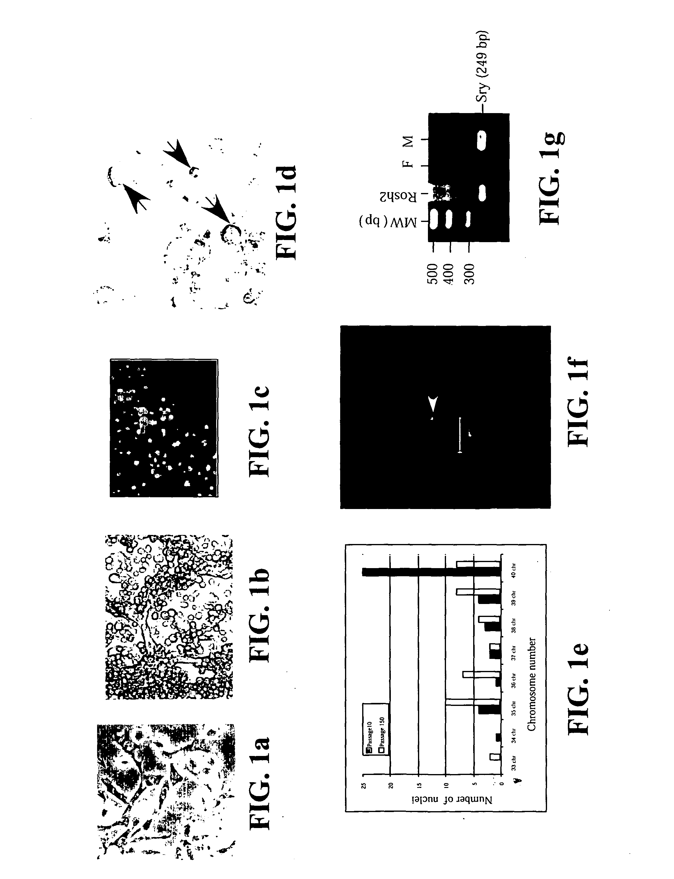
[0120] Preparation of Hemangioblast Cell Lines from Embryonic Stem Cells

[0121] 2.times.10.sup.4 single ES cells in 100 .mu.l ES media ES cells were cultured in 3.9 ml methycellulose media (MethoCult M3134, StemCell Technologies, Inc, Vancouver, Canada), 4.2 ml IMDM (Life Technologies, Rockville, Md.), 1.5 ml Serum, 100 .mu.l monothioglycerol stock solution (37.8 .mu.l in 10 ml PBS) (Sigma, St Louis, Mo.) 100 .mu.l 100X L-glutamine / Penicillin / Streptomycin stock solution (Life Technologies, Rockville, Md.). Six days later, colonies of cells or EBs were clearly visible to the naked eyes. The EBs were then dissociated into cell suspensions by incubating the EBs in 0.15% (w / v) collagenase / PBS supplemented with 20% (v / v) FCS at 37.degree. C. for 30 minutes and then disrupting the cell clumps by passing the solution through a syringe with a 20 -gauge needle 3 times. After another 30 minutes of incubation, the disruption was repeated with a 25-gauge needle. These cells were then plated on m...

example 3

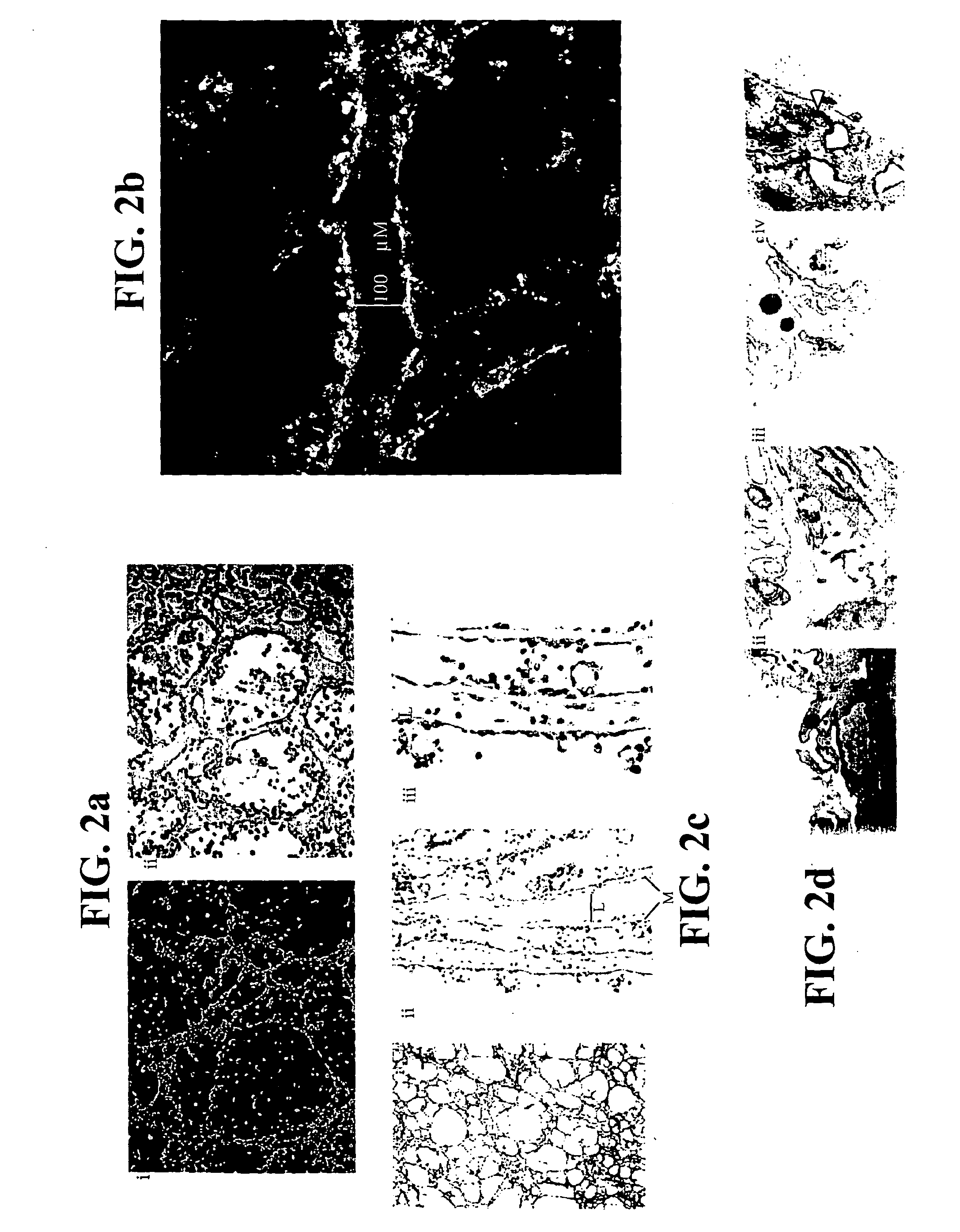
[0123] Preparation of Hemangioblast Cell Lines from Bone Marrow

[0124] Adult bone marrow (BM) was prepared from mice, pigs and humans. For mice, BM was flushed from the femurs of B6.129S7-GtRosa26 with saline using a needle and syringe. In pigs, BM was aspirated from the femur of pigs. Human BM was harvested by scraping from the split sternum of patients undergoing CABG surgery at NUH. The common denominator in all these procedures is the preservation of some BM tissue integrity in tissue clumps of 0.1 to 1 mm.sup.3 in volume. Each piece of tissue was cultured individually on 48-well mitomycin C-treated mouse embryonic fibroblast feeder plates in ES cell media. Most of the BM pieces attached to the plates within 24 hours. The cultures were maintained with changes of fresh media every two to four days. Over a period of one week, cells appeared to migrate out of the BM pieces. During the first week, the culture was a complex mix of cell types with much cell proliferation and cell death...

example 4

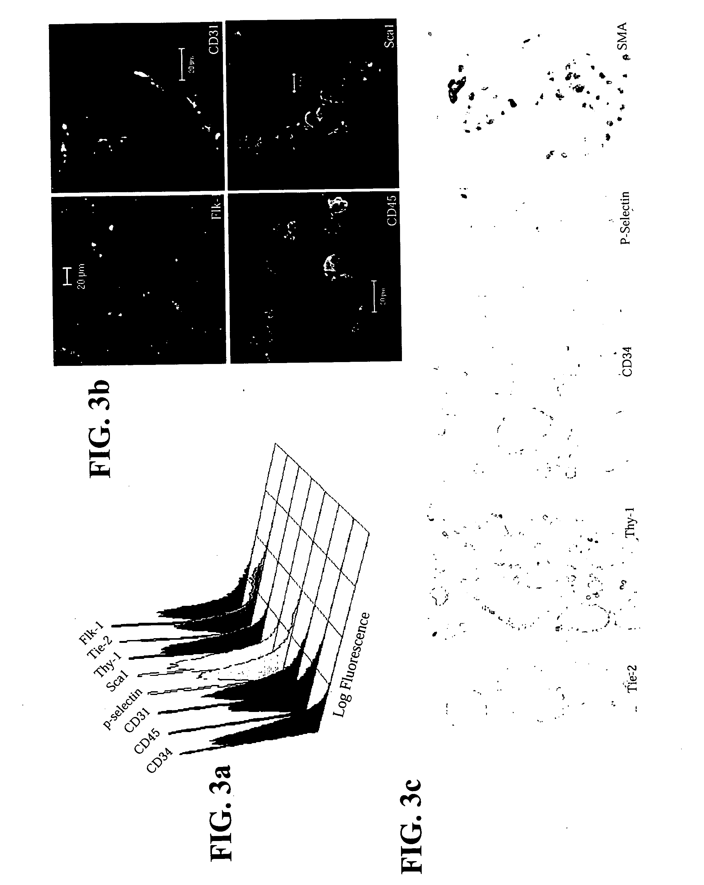
[0129] In vitro Differentiation

[0130] Like RoSH2 cells, Ro(BM)SH and HuSH cells can be induced to differentiate to form a mesh of tubular structures by plating at a density of 1.times.10.sup.6 cells on 6-cm tissue plates that were thinly coated with matrigel.


PUM

PropertyMeasurementUnit
population doubling timeaaaaaaaaaa
sizeaaaaaaaaaa

Abstract

The invention relates to isolated hemangioblast cells. Hematopoietic and endothelial cells are postulated to be derived from a common progenitor, hemangioblast. While hemangioblast has been isolated retrospectively during embryonic stem cell differentiation, it has not been isolated from embryos or from bone marrow. Prospectively stable clonal cell lines have been isolated from mammalian embryos, from embryonic stem cells and from mammalian bone marrow that can differentiate in vitro into tubular structures with both endothelial and hematopoietic markers such as CD34, CD31, Flk-1, TIE2, P-selectin, Sca-1, thy-1, CD45, and smooth muscle actin. Gene expression profiles in the undifferentiated and differentiated cells were consistent with endothelial and hematopoietic differentiation potential. Transplantation studies in isogenic or immunodeficient mice demonstrated that these cells were not tumorigenic. In an appropriate microenvironment, the cells incorporate into the vasculature and participate in hematopoiesis.

Description

[0001] The present invention relates to the derivation of hemangioblast cell lines which have the potential to differentiate into hematopoietic and endothelial cells in vitro and in vivo.BACKGROUND OF THE DISCLOSURE[0002] Hematopoiesis and vasculogenesis are closely associated events that develop in tandem spatially and temporally during embryogenesis (Murray, 1932; Sabin, 1920). Primitive hematopoiesis and the establishment of the yolk sac vasculature occur simultaneously when mesodermal cells in the presumptive yolk sac proliferate and differentiate to form vascular structures with primitive erythroblasts known collectively as blood islands. Hematopoiesis during mouse development is well characterized (Keller et al., 1999). Blood islands are visible in the yolk sac at 7.5 days post coitus (dpc). By 11.5 dpc, the fetal liver displaces the yolk sac as the major site of hematopoiesis in mouse embryo and also signifies the switchover to definitive hematopoiesis. Unlike primitive hemat...

Claims


Application Information

Patent Timeline
Patent Type & Authority Applications(United States)
IPC IPC(8): A61K35/12C12N5/074
CPCA61K2035/124C12N2506/02C12N5/0692
Inventor LIM, SAI KIANG
Owner NAT UNIV OF SINGAPORE
Why Eureka
Try Eureka