
Tissue implantable sensors for measurement of blood solutes

a technology of blood solutes and implantable sensors, which is applied in the field of tissue implantable sensors for blood solute measurement, can solve the problems of impaired blood glucose regulation, near inability to sufficiently frequent sampling, and obstruct warning strategies that rely on, so as to improve the results of estimation, the effect of easing the effects of intrinsic detector drift and failur

Active Publication Date: 2005-03-17
RGT UNIV OF CALIFORNIA
View PDF21 Cites 133 Cited by
  • Summary
  • Abstract
  • Description
  • Claims
  • Application Information

AI Technical Summary

Benefits of technology

Systematically combining and adjusting data obtained from multiple detectors. (primary, secondary or both) in a sensor according to preselected and calibrated signal adjustment coefficients compensates for inaccuracies in measurements caused by point-to-point variations in analyte concentration and in the magnitude of confounding phenomena, as well as variations over time in same. Such variations may result from variations in tissue vascularization (either within the same tissue over time, or between biological microenvironments in a given tissue at a given time, or between implantation sites); changes in local or regional blood flow at the implantation site; variations in microvascular activity or vasomotion, or formation of fibrous tissue over portions of the implanted sensor. The method of systematically combining and adjusting signals of the present invention also provides a system for ameliorating effects of intrinsic detector drift and failure.

Problems solved by technology

Regulation of blood glucose is impaired in diabetes by the inability of the pancreas to adequately produce the glucose-regulating hormone insulin and by the insensitivity of various tissues that use insulin to take up glucose.
This procedure has several disadvantages, including: (1) the discomfort associated with fingersticking, which should be performed repeatedly each day; (2) the near impossibility of sufficiently frequent sampling (some blood glucose excursions require sampling every 20 minutes, or less, to accurately treat); and (3) the requirement that the user initiate blood collection, which precludes warning strategies that rely on automatic early detection.
Using the present procedure, the frequent sampling regimen that would be most medically beneficial cannot be realistically expected of even the -most committed patients, and -automatic sampling, which would be especially useful during periods of sleep, is not available.
Although this sensor provided a continuous recording of blood glucose, which is most advantageous for clinical applications, implantation at a central venous site poses risks of blood clot formation and vascular wall damage.
One problem that has confronted previous attempts to implant sensors in solid tissue is that the pattern of blood vessels in the vicinity of the sensor may be highly variable, and may change with time in response to the implantation procedure and the presence of an implant.
In other cases, blood vessels are more distant and sensors appear not to function, to function weakly, or to function only with substantial delays.
The occurrence of such complete occlusion is, of course, not predictable.
Although using biocompatible materials can minimize the tissue response to the implant, capillary distributions or diffusion resistances may still be affected, altering the diffusive flux to the sensor.

Method used


Image

  • Tissue implantable sensors for measurement of blood solutes
  • Tissue implantable sensors for measurement of blood solutes
  • Tissue implantable sensors for measurement of blood solutes

Examples


example 1

Method for Fabricating a Tissue Implantable Glucose Sensor

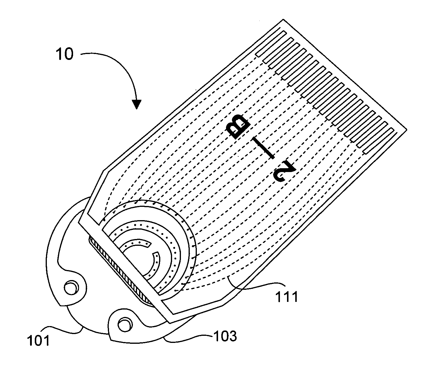
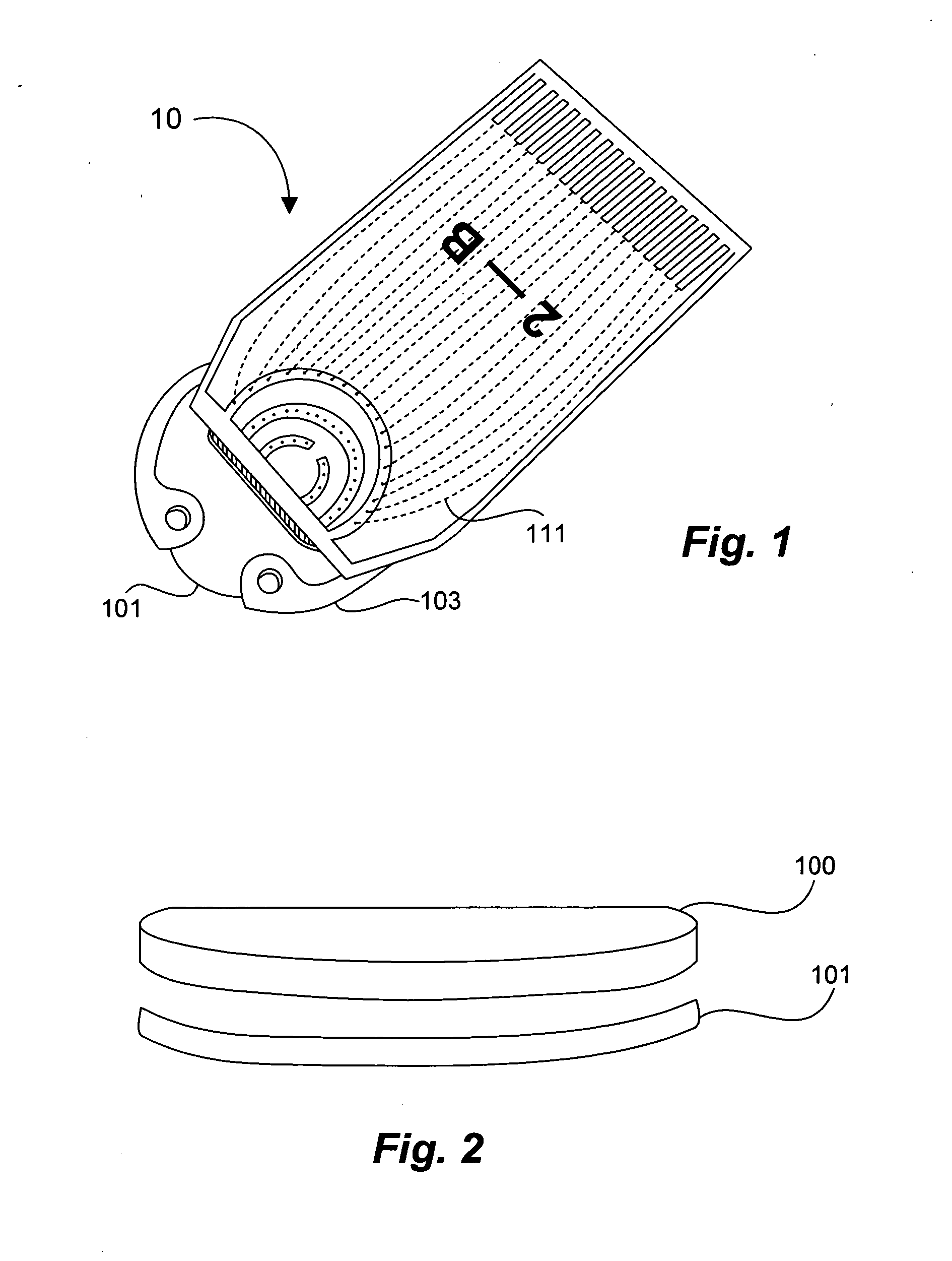
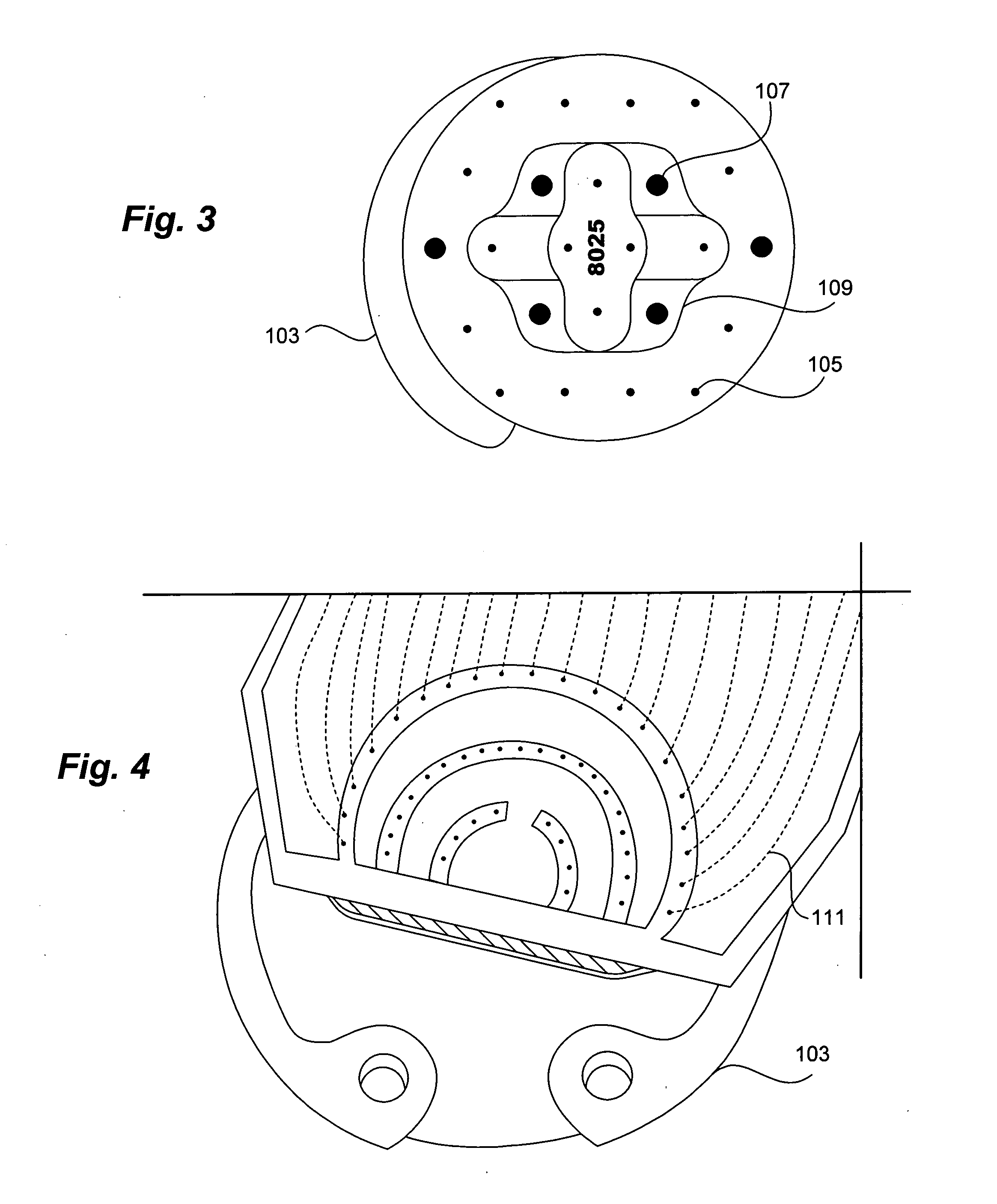
1) Using standard thick-film techniques, create conductive vias through an implant-grade alumina disc. 2) Print and fire an electrode array of high-purity platinum onto the disc, aligning the electrodes with their respective conductive vias. These electrodes will become the working, reference and counter electrodes of the detectors. 3) Print and fire insulating dielectric layers, as necessary. 4) Electrochemically apply Ag / AgCl to the reference electrodes. 5) Using wirebonding, or other conventional circuit fabrication techniques, provide connections from the electrodes of the detector array to conventional potentiostat circuitry. 6) Affix a multilayer glucose oxidase-impregnated membrane to the face of the disc. The outer layer of this membrane is silicone rubber; the glucose oxidase is impregnated in a fashion that supplies oxygen in relative excess to the glucose oxidase (see, for example, co-pending and commonly ow...

example 2

Improvement in Stability of Glucose Measurements Using the Sensor of the Invention

FIG. 5 depicts data typical of current readings obtainable in vitro from a dual-detector sensor, consisting of a glucose-sensitive detector and an oxygen-sensitive detector.

A sensor constructed as described in Example 1 was implanted in a live, healthy hamster. In particular, two titanium plates, each of which includes a small “window,” are placed so as to support a thin layer of retractor muscle beneath excised subcutaneous tissue on the animal's back. A cover glass was placed in the window over one side of the exposed skin, and the sensor device was fixed onto the opposite side. Catheters were placed in the carotid artery and jugular vein for sampling and fluid delivery. The resulting structure is a layer of tissue 100 μm thick by 12 mm in diameter, having intact microvasculature.

To provide stimulus to the glucose detector, a bolus of 60 μl / minute of 50 gm % glucose was introduced into the cathe...

example 3

Application of Signal Processing Algorithms

In the following figures, the time course of measurements taken is indicated along the horizontal axis of each graph. Either the measured current or detected analyte concentration is indicated along the vertical axis of each graph, as shown.

FIG. 11 shows unprocessed primary and secondary detector signals from a glucose sensor that uses oxygen detectors and the glucose oxidase-catalyzed depletion of oxygen to measure glucose, the sensor being made according to the invention. The perturbation at about 12:25 in each of the primary detector signals is approximately simultaneous with the others, but not simultaneous with the secondary detector signals. Thus, the primary detectors appear to be responding approximately in synchrony with each other.

FIG. 12 shows an enlarged view of the secondary detector signals during a time period where a perturbation has been detected in the signals of both the primary and secondary detectors due to a change...


PUM


Abstract

A tissue-implantable sensor for measurement of solutes in fluids and gases, such as oxygen and glucose, is provided. The sensor includes a multiplicity of detectors, constructed and arranged to improve the probability that one or more detectors will have access to a vascular source at points in time sufficient to permit accurate measurements to be taken. Means and methods for calculating solute levels using the sensor device of the invention are also provided.

Description

BACKGROUND OF THE INVENTION Regulation of blood glucose is impaired in diabetes by the inability of the pancreas to adequately produce the glucose-regulating hormone insulin and by the insensitivity of various tissues that use insulin to take up glucose. To correct this disregulation requires blood glucose monitoring. Currently, glucose monitoring in the diabetic population is based largely on collecting blood by “fingersticking” and determining its glucose concentration by conventional assay. This procedure has several disadvantages, including: (1) the discomfort associated with fingersticking, which should be performed repeatedly each day; (2) the near impossibility of sufficiently frequent sampling (some blood glucose excursions require sampling every 20 minutes, or less, to accurately treat); and (3) the requirement that the user initiate blood collection, which precludes warning strategies that rely on automatic early detection. Using the present procedure, the frequent sampl...

Claims


Application Information

Patent Timeline
Patent Type & Authority Applications(United States)
IPC IPC(8): A61B5/00
CPCA61B5/0031A61B5/14865A61B5/14532
Inventor GOUGH, DAVID A.JABLECKI, MICHAEL C.LUCISANO, JOSEPH Y.CATLIN, MARK B.
Owner RGT UNIV OF CALIFORNIA
Learn More