Application of electrical stimulation for functional tissue engineering in vitro and in vivo
a technology of functional tissue engineering and electrical stimulation, which is applied in the direction of skeletal/connective tissue cells, prosthesis, therapy, etc., can solve the problem of biocompatibility of the substrate used in the preparative methods, and achieve the effect of enhancing the integration of engineered and host tissues
- Summary
- Abstract
- Description
- Claims
- Application Information
AI Technical Summary
Benefits of technology
Problems solved by technology
Method used
Image
Examples
example 1
Preparation of Bioartiflcial Cardiac Muscle Tissue
[0153] Cardiac Myocyte Preparation. Primary cultures of cardiac myocytes were prepared by enzymatic digestion of ventricles obtained from neonatal (2 day old) Sprague-Dawley rats (Taconic), as previously described (M. Toraason et al., Toxicology, 1989, 56: 107-113, the contents of which are incorporated herein by reference). Briefly, ventricles (n=50, 5 litters in 3 independent studies) were incubated with 0.1% trypsin overnight and dissociated in four to five sequential steps using 0.1% collagenase.
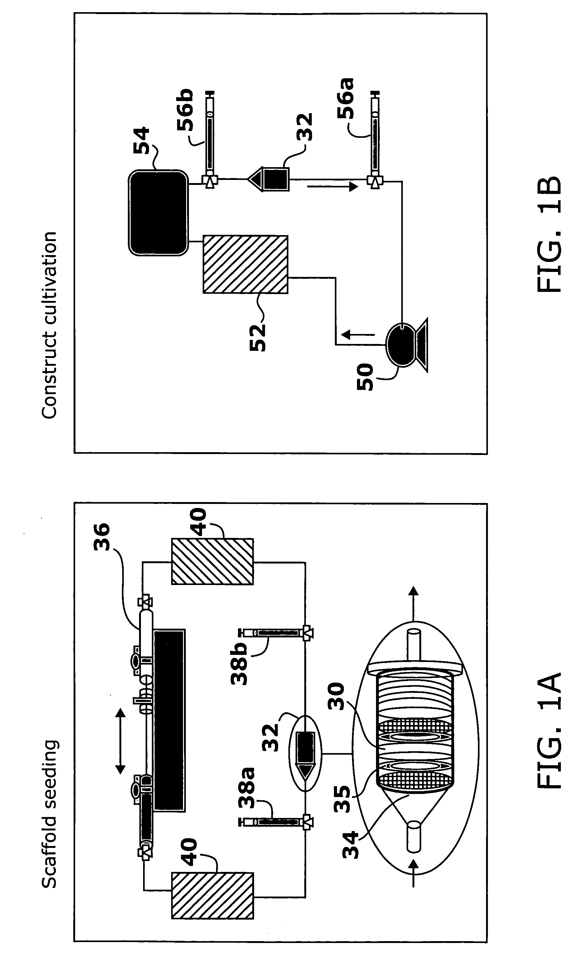
[0154] Cell-Seeded Construct Preparation. Isolated cardiac myocytes were suspended in Matrigel® (106 cells / 5 μL) and seeded onto Ultrafoam™ collagen sponge scaffold (6×8×1.5 mm) at the density of 0.8×10⁸ cells / cm3.
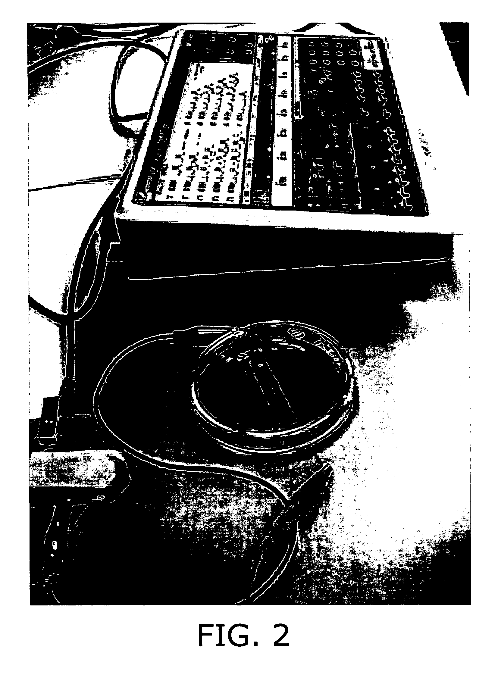
[0155] Cultivation of Cell-Seeded Construct. After 3 days of cultivation, the engineered tissue was transferred to a chamber consisting of a 100 mm glass Petri dish fitted with two ¼″ diameter carbon rods (stimulating electro...
example 2
Histological and Immunohistochemical Assessments
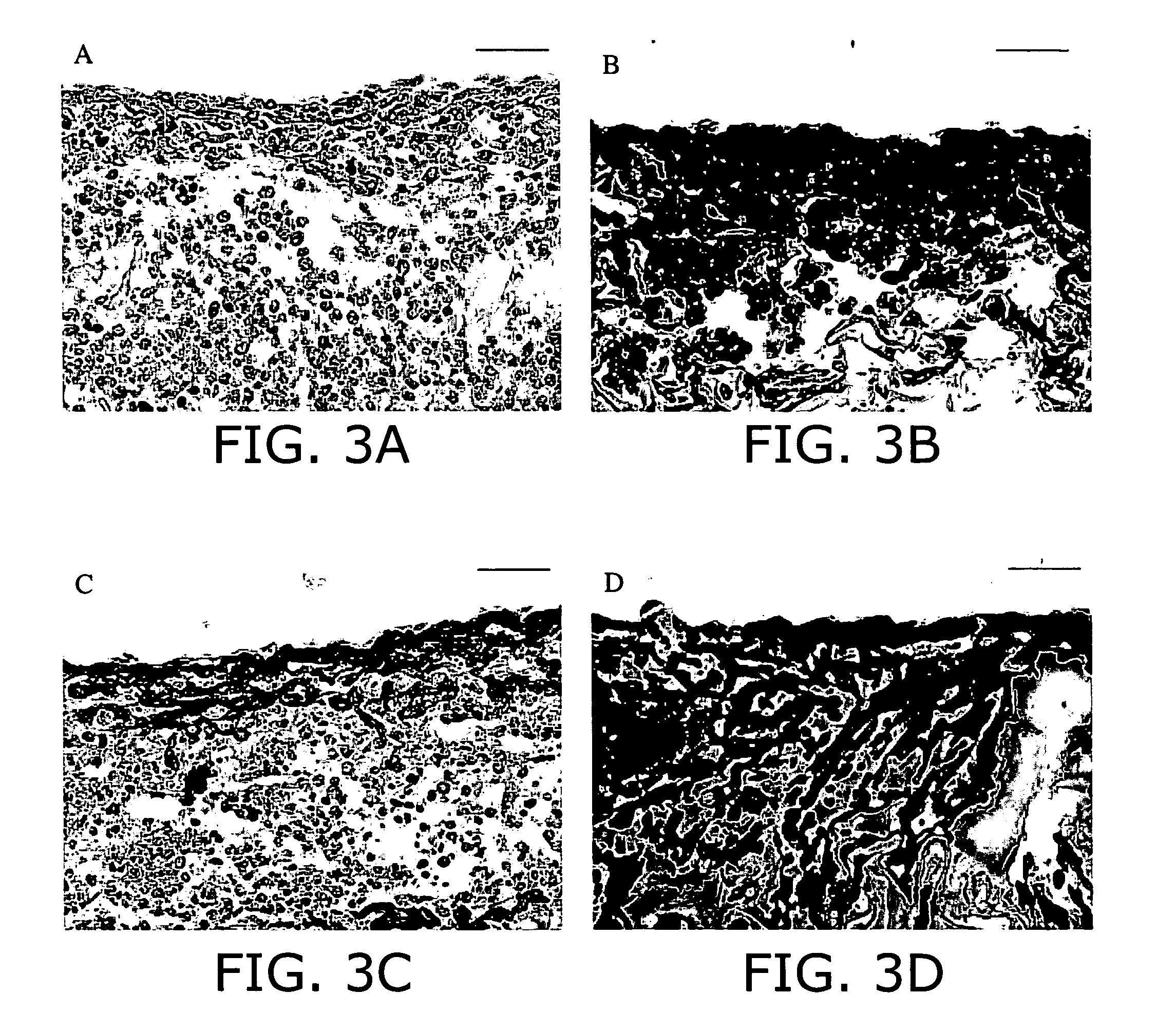
[0157] General Evaluation. Cardiac muscle constructs obtained as described in Example 1 in the presence or in the absence of electrical stimulation were rinsed in PBS, and immersed in 10% neutral buffered formalin (Sigma-Aldrich). Samples were embedded in paraffin, sectioned at 5 μm, and stained with hematoxylin and eosin (H+E) for general evaluation.
[0158] Tissue Architecture and Cell Distribution. Staining for cardiac troponin I, sarcomeric α-actin, α-myosin heavy chain (α-MHC), β-myosin heavy chain (β-MHC) (myocyte-specific contractile proteins) and connexin-43 (Cx-43, gap junctional protein) was used to assess the fraction and distribution of cardiac myocytes in the constructs.
[0159] For immunohistochemical staining, cardiac tissue sections were depariffinized and antigen was retrieved by heat treatment for 20 minutes at 95° C. in decloaking chamber (Biocare Medical). Subsequently, endogenous peroxidase activity was quenched by ...
example 3
Biochemical and Molecular Assays
[0163] DNA and Protein Assays. Samples for DNA, total protein, and Western blot analyses are homogenized in buffer (1 M NH4OH / 2% Triton X-100, 0.04 mL / mg wet wt of sample) for 1 minute. DNA is measured fluorimetrically by Hoescht binding, and total protein is measured by a commercially available kit (Bio-Rad) as previously described (N. Bursac et al., Am. J. Physiol. Heart Circ. Physiol. 1999, 277: H433-H444; R. L. Carrier et al., Biotechnol. Bioeng. 1999, 64: 580-589).
[0164] Protein Analysis. The protein expression levels of creatine kinase-MM (CK-MM) and myosin heavy chain (MHC), troponin I and connexin-43 in constructs are quantified as markers of cellular differentiation. Creatine kinase, a dimer of M- or B-type subunits, plays a vital role in the maintenance of cytosolic ATP (L. H. Opie, The Heart Physiologyfrom Cell to Circulation, L. Opie (Ed.), Lippincott-Raven: Philadelphia, 1998, pp. 295-342), whereas MHC is involved in the generation of c...
PUM
 Abstract
Description
Claims
Application Information
 - R&D
- Intellectual Property
- Life Sciences
- Materials
- Tech Scout
- Unparalleled Data Quality
- Higher Quality Content
- 60% Fewer Hallucinations
Browse by: Latest US Patents, China's latest patents, Technical Efficacy Thesaurus, Application Domain, Technology Topic, Popular Technical Reports.
© 2025 PatSnap. All rights reserved.Legal|Privacy policy|Modern Slavery Act Transparency Statement|Sitemap|About US| Contact US: help@patsnap.com


