[0022] There is a great clinical need for the treatment of glaucoma by a method that is faster,
safer, and less expensive than currently available modalities, and by implanting a device having
pressure sensing capability for transporting aqueous from the anterior chamber to Schlemm's canal.
[0023] Glaucoma surgical morbidity would greatly decrease if one were to bypass the focal resistance to outflow of aqueous only at the point of resistance, and to utilize remaining, healthy aqueous outflow mechanisms. This is in part because episcleral
aqueous humor exerts a backpressure that prevents intraocular pressure from going too low, and one could thereby avoid hypotony. Thus, such a
surgery would virtually eliminate the risk of hypotony-related
maculopathy and choroidal hemorrhage. Furthermore, visual
recovery would be very rapid, and the
risk of infection would be very small, reflecting a reduction in incidence from 2-5% to about 0.05%.
[0024] Techniques performed in accordance with embodiments herein may be referred to generally as “trabecular
bypass surgery.” Advantages of the present invention include lowering intraocular pressure in a manner which is simple, effective,
disease site-specific, and can potentially be performed on an outpatient basis.
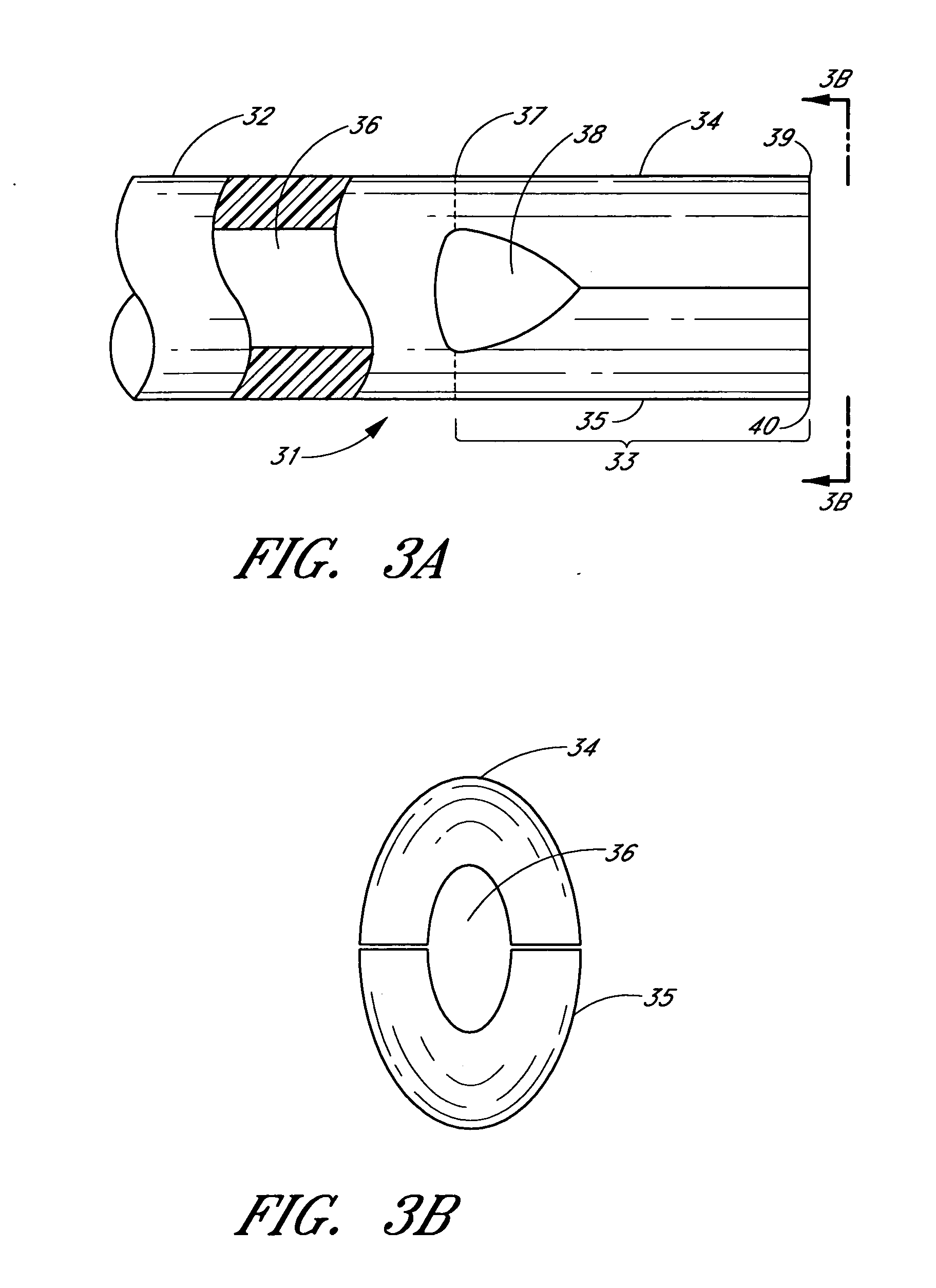
[0031] In one embodiment, at least one of the two bifurcatable elements has a tapered distal end, adapted for
insertion ease. The trabecular shunt may have its surface coated with a
coating material selected from one or more of the following:
polytetrafluoroethylene (e.g., Teflon™),
polyimide, hydrogel,
heparin, hydrophilic compound, anti-angiogenic factor, anti-proliferative factor, therapeutic drugs, and the like. The
surface coating material may also provide a mechanism for site-specific therapies.
[0032] In one embodiment, the device of the invention may include a flow-restricting member for restricting at least one component in fluid. The flow-restricting member may be a filter comprising one or more
filtration materials selected from the following:
expanded polytetrafluoroethylene,
cellulose,
ceramic, glass, Nylon, plastic, fluorinated material, or the like. The flow-restricting member may advantageously be a filter selected from the following group of filter types: hydrophobic, hydrophilic, membrane, microporous, and non-woven. The flow-restricting member acts to limit or prevent the
reflux of any undesired component or contaminant of blood, such as red blood cells or
serum protein, from the aqueous veins into the anterior chamber. It is useful to
restrict one or more of the following components or contaminants: platelets, red blood cells, white blood cells, viruses,
bacteria, antigens, and toxins.
[0046] Among the advantages of trabecular
bypass surgery in accordance with the invention is its simplicity. The
microsurgery may potentially be performed on an outpatient basis with rapid visual
recovery and greatly decreased morbidity. There is a
lower risk of infection and choroidal hemorrhage, and there is a faster
recovery, than with previous techniques.

