Methods for mass spectrometry detection and quantification of specific target proteins in complex biological samples

a mass spectrometry and target protein technology, applied in the field of mass spectrometry detection and quantification of specific target proteins in complex biological samples, can solve the problems of difficult to obtain accurate quantitative information concerning the levels of identified proteins and the levels of site-specific or other modifications of individual protein molecules, and the development of an elisa for a target protein is a particularly laborious and lengthy task, and achieves rapid and sensitive detection, the effect of increasing the reliability of the diagnostic results obtained

Inactive Publication Date: 2005-07-14
ISTA
View PDF8 Cites 38 Cited by
  • Summary
  • Abstract
  • Description
  • Claims
  • Application Information

AI Technical Summary

Benefits of technology

[0046] The present method for overcomes limitations associated with mass spectrometry sytems that quantify only relative concentrations by ICATs, N-succinylation or other mass spectrometry methods that involve chemical modification of the parent protein because it does not require the presence of specific amino acids that are accessible to reactive agents, or a second protein mixture for quantification. The present invention provides for multiple, simultaneous, independent analyses of the protein of interest, thereby significantly increasing the reliability of the diagnostic results obtained.
[0047] Beyond the foregoing, the invention described can be employed for the quantitative analysis of targeted proteins in complex biological...

Problems solved by technology

Despite these advances, it remains difficult to obtain accurate quantitative information concerning the levels of the identified proteins and the levels of site-specific or other modifications to individual protein molecules.
The development of an ELISA for a target protein is a particularly laborious and lengthy task, which requires production of monospecific antibodies to one or more epitopes residing within the protein.
In addition, the occurrence of false positives and negatives is not uncommon.
Thus, they may have insufficient sensitivity for commercial diagnoses, particularly in cases where there is significant risk of legal liability in the event of an incorrect result.
Further, ELISA-based assays are limited in that they can only detect analytes for which antibodies have been raised.
This requires prior knowledge of sample composition coupled with time-consuming effort in order to prepare sufficient purified protein and raise a new antibody for each target pr...

Method used


Image

  • Methods for mass spectrometry detection and quantification of specific target proteins in complex biological samples
  • Methods for mass spectrometry detection and quantification of specific target proteins in complex biological samples
  • Methods for mass spectrometry detection and quantification of specific target proteins in complex biological samples

Examples


example 1

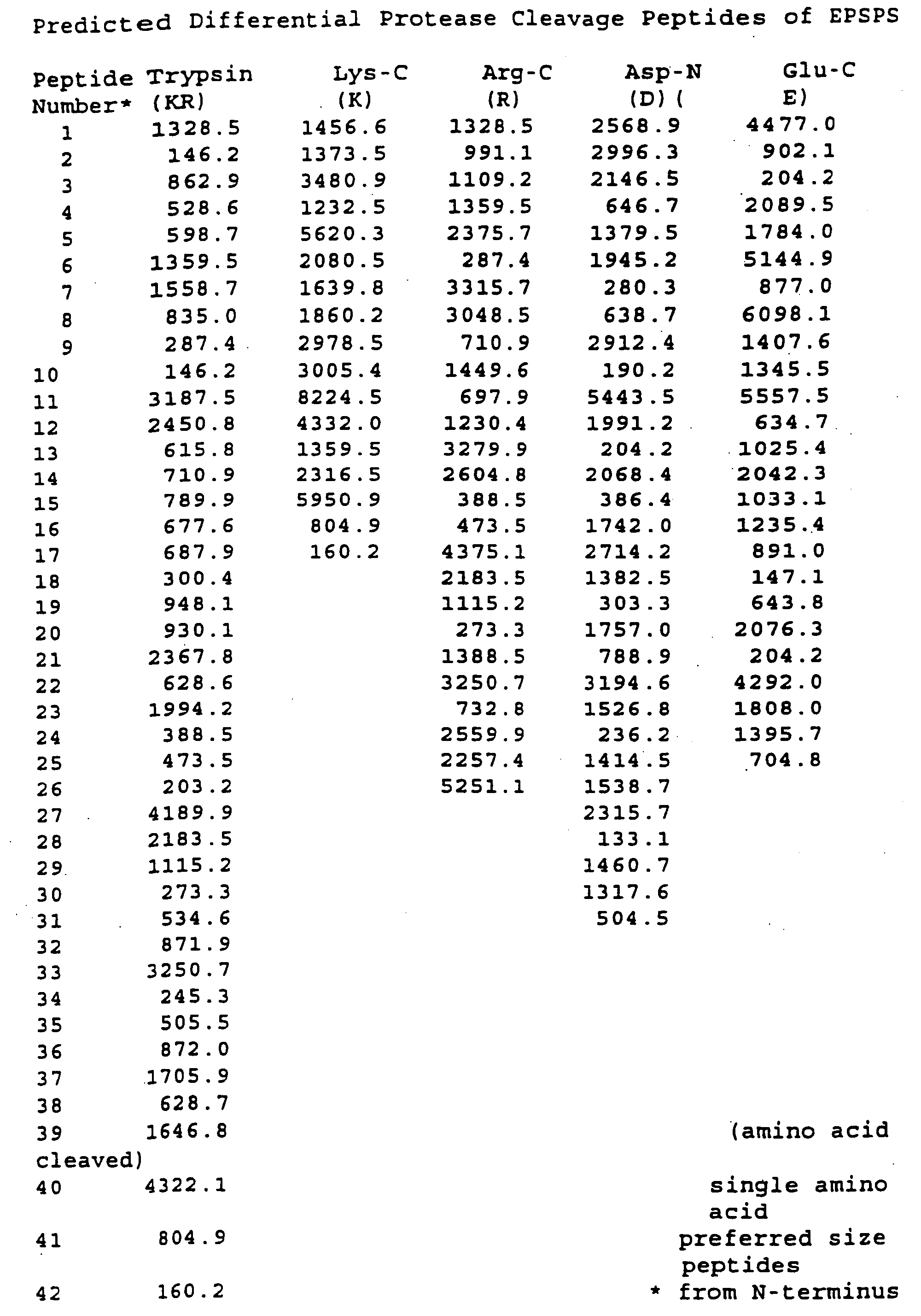
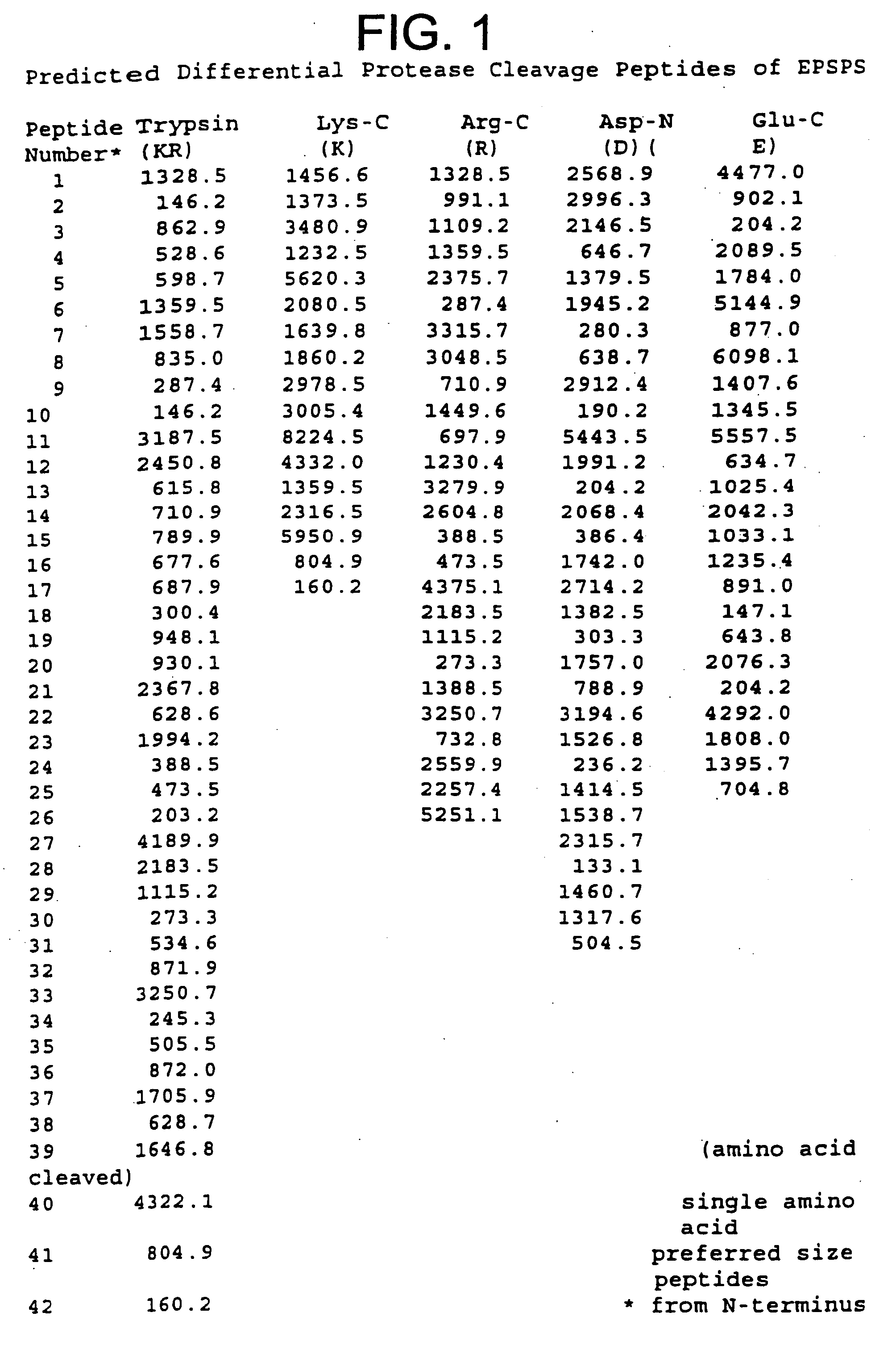
Predicted Proteolytic Cleaveage of 5-enolpyruvylshikimate-3-phosphate Synthase Present in Genetically Modified Plants

[0091] For those proteins that correspond to genes that have been cloned and sequenced, such as for 5-enolpyruvylshikimate-3-phosphate synthase (EPSPS), its amino acid sequence can be readily deduced from the DNA sequence. The deduced amino acid sequence of EPSPS was subjected to theoretical or in silico protease digestion using 5 distinct proteases. Three of the proteases cleave at basic amino acid sites (trypsin, Arg-C, Lys-C) and the two other proteases at acidic sites (Asp-N and Glu-C). Arg-C and Lys-C cleave at the carboxy-terminus of arginine and lysine residues, respectively. Trypsin cleaves at two amino acid sites, lysine and arginine residues. Asp-N cleaves at amino-terminus of aspartic residues while Glu-C cleaves at the carboxy-terminus of glutamic residues.

[0092] In FIG. 1, the peptides cleaved by the five proteases are listed from the amino-terminus to ...

example 2

Enrichment of 5-enolpyruvylshikimate-3-phosphate Synthase EPSPS from Soybean Seeds

[0093] Enrichment of 5-enolpyruvylshikimate-3-phosphate synthase from transgenic soybean seeds.

[0094] The initial step for the detection and quantitation of a GM protein in plants is to obtain a protease digest profile of peptides for the protein of interest. The protein may be available in large amounts from a recombinant source such as bacteria or insect (baculovirus expressed) cells. Alternatively, transgenic plants expressing sufficient levels of the protein may provide the material required for peptide profiling. For EPSPS, the latter source was used as the source for peptide profiling.

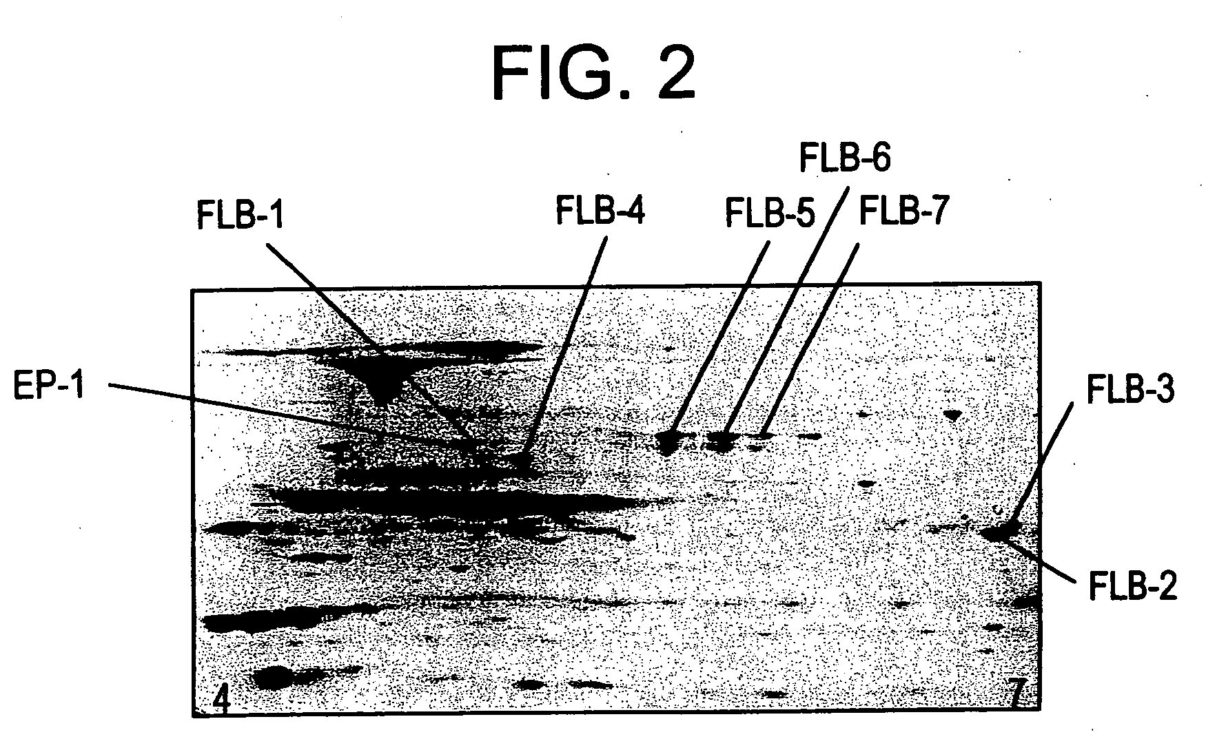
[0095] The plant material was subjected to a series of enrichment protocols for the identification of EPSPS and was preliminarily identified when comparing colloidal stained 2-D gel patterns of proteins isolated from non-transformed and transgenic soybean seeds expressing EPSPS.

[0096] Ten seeds were ground in a ...

example 3

Identification of Diagnostic Marker Peptides for 5-enol-pyruvylshikimate-3-phosphate Synthase and beta-conglycin in Soybean Seeds

[0104] Identification of EP-1 and FLB-1 FLB-7 diagnostic protein markers.

[0105] The eight protein spots detectable on a 2-D gel corresponding to EP-1 and FLB-1 through FLB-7 were excised, proteolytically cleaved, analyzed and identified as described below.

[0106] Identical protein spots from 1-3 gels were excised manually and pooled. The gel pieces were macerated and destained with 25 mM ammonium bicarbonate / 50% acetonitrile in a 1.5 mL microfuge tube with vigorous shaking for 30 minutes. The blue-tinted destaining solution was removed and discarded with a fine-tip pipette. The destaining step was repeated until the stain was removed from the gel pieces. The gel pieces were dried under vacuum for 10 to 15 minutes. Proteins were digested overnight at 37° C. in a total volume of 25 μL of sequence-grade, modified trypsin (Roche Diagnostics) at a final prote...


PUM


Abstract

Mass spectrometry-based methods are described for the detection or quantification of targeted proteins in biological samples e.g., plants, animals, and microorganisms, parts (e.g., tissue or cells) thereof, and products derived from plants, animals or microorganisms.

Description

CROSS REFERENCE TO RELATED APPLICATIONS [0001] This application a continuation of U.S. application Ser. No. 10 / 475,206 filed Oct. 17, 2003, which is a national phase entry under 35 U.S.C. § 371 of International Application PCT / US02 / 12014, filed Apr. 17, 2002, published in English, which claims benefit of U.S. Provisional Patent Application 60 / 284,273, filed Apr. 17, 2001, and U.S. Provisional Patent Application 60 / 284,713, filed Apr. 18, 2001. The disclosures of all of said applications are incorporated by reference herein.TECHNICAL FIELD [0002] The present invention relates generally to the use of mass spectrometry to detect, quantify, and characterize target proteins in complex biological samples. BACKGROUND OF THE INVENTION [0003] The development of sensitive, precise, high throughput methods for directly monitoring specific proteins is increasingly desirable for diagnostic applications in both agricultural and clinical fields. Recent advances in the use of mass spectrometry (MS)...

Claims


Application Information

Patent Timeline
IPC IPC(8): C12Q1/68G01N33/53G01N33/537G01N33/543G01N33/68
CPCG01N33/6848
Inventor EVERETT, NICHOLASPETELL, JAMESYOUNG, SCOTT
Owner ISTA
Why Eureka
Try Eureka