Decellularized grafts from umbilical cord vessels and process for preparing and using same

Inactive Publication Date: 2005-09-15
THE BOARD OF RGT UNIV OF OKLAHOMA
View PDF15 Cites 83 Cited by
  • Summary
  • Abstract
  • Description
  • Claims
  • Application Information

AI Technical Summary

Benefits of technology

[0038]FIG. 6 graphically illustrates the burst pressures of the aHUV compared to mHUV and decellularized HUV (dHUV). The burst pressure of aHUV was significantly higher than that of the mHUV. There is a dramatic drop in both range and variance associated with automated dissection.
[0039]FIG. 7 graphic

Problems solved by technology

Current treatments are limited to reconstructive surgery where a patient's own (autologous) non-diseased vessels are relocated to bypass diseased or blocked blood vessels that would ultimately result in cardiac arrest, stroke or death.
Maintaining the flow of oxygen-rich blood to organs down-stream of severely occluded blood vessels is often limited to surgically by-passing diseased sections with substitute vessels.
Small diameter (<6 mm) vascular reconstructions have proven considerably more problematic than large diameter vessel reconstructions, where reduced diameter and low flow rates, compounded by high resistance, amplify poor biological host/graft interactions.
Autologous arterial grafts are preferred over synthetics because the latter fail due to poor biocompatibility and/or mechanical properties.
However, there are several structural limitations when the umbilical cord vein is used as a glutaraldehyde ‘fixed’ material.
Although cryo-preserved veins are a promising option, they too are limited by supply (Dresdale et al., 1990; Fujitani et al, 1992; and Davies, 1994), and umbilical cord veins, in their current form, have not proven reliable due (primarily) to a potential for aneurysm formation.
The reduced immune response attributed to cross-linking has been described as a ‘masking’ of allogenic or xenogenic components that would otherwise be seen as foreign, resulting in chronic rejection and subsequent failure of the graft.
The quandary is that chemical treatments designed to stabilize an

Method used


Image

  • Decellularized grafts from umbilical cord vessels and process for preparing and using same
  • Decellularized grafts from umbilical cord vessels and process for preparing and using same
  • Decellularized grafts from umbilical cord vessels and process for preparing and using same

Examples


example 1

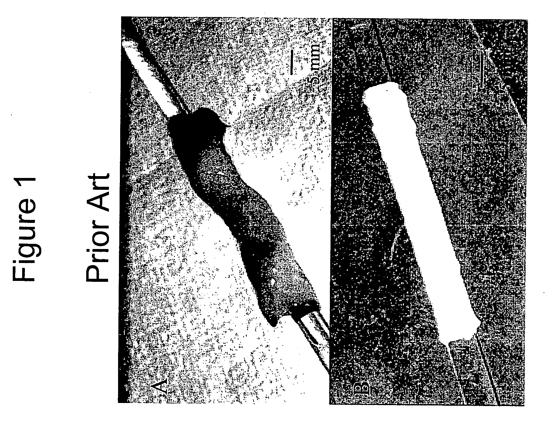
[0090]FIG. 1 illustrates a whole human umbilical cord (FIG. 1A), and an umbilical vein obtained from the umbilical cord by a manual dissection procedure of the prior art (FIG. 1B). Typically, manual dissection methods require about one to about three hours to produce one viable vessel, and extensive mechanical variation is displayed across the manually dissected vein. These tedious and error prone dissection methods of the prior art have restricted the veins application, unless the vessel is extensively cross-linked and reinforced with synthetic polymers (Dardik et al., 1988; and Dardik, 1995).

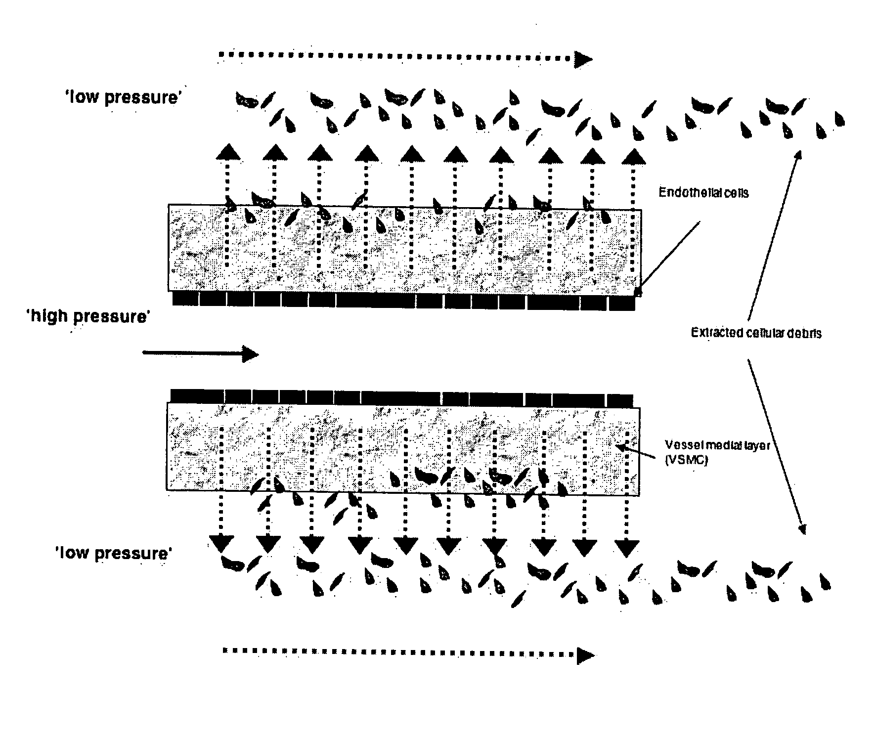
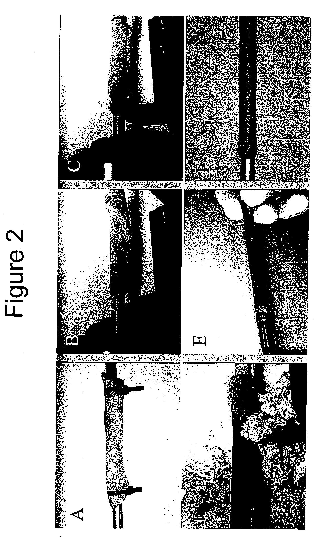
[0091]FIG. 2 illustrates the unique automated dissection method of the present invention. Using the automated dissection method of the present invention, an umbilical vein can be extracted from the umbilical cord in a maximum of about 2 minutes, producing a mechanically uniform material (Daniel et al., 2004; McFetridge et al., 2004; and Daniel et al., 2004). Briefly, the method of the present...

example 2

[0106] By progressively modifying cell culture conditions to mimic the environment an implanted construct might encounter in vivo, the effect of mechanical force and hypoxia on hVSMC proliferation, migration, and remodeling processes is assessed. In order to quantify these distinct environmental conditions, three areas are investigated: (1) traditional cell culture systems, (2) introduction of mechanical stress, and (3) exposure to hypoxic conditions. Variation between the three areas is quantified using standardized experimental and analytical methods.

[0107] First, the ability of the HUV bioscaffold to provide a favorable environment for early regenerative events is assessed. To quantify the regenerative capacity of the decellularized HUV bioscaffold under traditional ‘static’ tissue culture conditions using primary human SMC is investigated. Cell proliferation and viability using standard histological and immunohistochemical techniques is investigated. Image analysis software ass...

example 3

[0113] In another embodiment of the present invention, the tissue graft of the present invention is utilized for the development of a multi-functional ex vivo acellular bioscaffold for oral wound repair. Due to structural and morphological variation between the upper (lumen) and lower (ablumen) surfaces of the vascular scaffold, the smooth, type IV collagen surface is disposed outermost, in contact with the oral cavity, and the more fibrous surface of type I collagen and hyaluronic acid is disposed directly on top of the wound site. These investigations examine the rate of cell re-population and remodeling of the scaffold from cells directly within the wound site. The second application of the present invention in oral wound repair is as a tissue engineered construct seeded with autologous cells, then implanted as functional tissue. The experiments described herein support both applications by gaining a thorough understanding of the material's ability to re-integrate with biological...


PUM


Abstract

A tissue graft is disclosed that includes a decellularized umbilical vessel having a luminal surface and an ablumenal surface, wherein the decellularized umbilical vessel is prepared by an automated dissection process. The decellularized umbilical vessel has not been substantially cross-linked. In one method of use, the decellularized umbilical vessel is capable of having at least one cell type seeded at least a portion of at least one of the luminal and ablumenal surfaces thereof. A process of preparing the tissue graft, methods of using the tissue graft, and kits which contain the tissue graft are also disclosed.

Description

CROSS-REFERENCE TO RELATED APPLICATIONS [0001] This application claims benefit under 35 U.S.C. 119(e) of U.S. provisional patent application Ser. No. 60 / 551,607, filed Mar. 9, 2004, entitled “TISSUE ENGINEERING VASCULAR GRAFTS USING THE UMBILICAL VEIN AS A DECELLULARIZED MATRIX AND A RAPID BIOREACTOR-BASED AUTOLOGOUS CELL SEEDING TECHNOLOGY FOR BYPASS SURGERY”, which is hereby expressly incorporated herein by reference in its entirety.STATEMENT REGARDING FEDERALLY SPONSORED RESEARCH OR DEVELOPMENT [0002] Not applicable. BACKGROUND OF THE INVENTION [0003] 1. Field of the Invention [0004] The present invention relates to decellularized grafts for tissue engineering, and more particularly but not by way of limitation, to decellularized grafts from umbilical cord vessels for tissue engineering as an acellular matrix and for use with cell seeding methodology. [0005] 2. Brief Description of the Art [0006] Vascular disease is the number one cause of death in Western societies. Current trea...

Claims


Application Information

Patent Timeline
IPC IPC(8): A61F2/02A61L27/36A61L27/38C12N5/071
CPCA61L27/3604A61L27/3683C12N2533/54C12N5/0691A61L27/3886A61L27/3625
Inventor MCFETRIDGE, PETER S.
Owner THE BOARD OF RGT UNIV OF OKLAHOMA
Why Eureka
Try Eureka