

Method for determining endoglycosidase enzyme activity
- Summary
- Abstract
- Description
- Claims
- Application Information
AI Technical Summary
Benefits of technology
Problems solved by technology
Method used
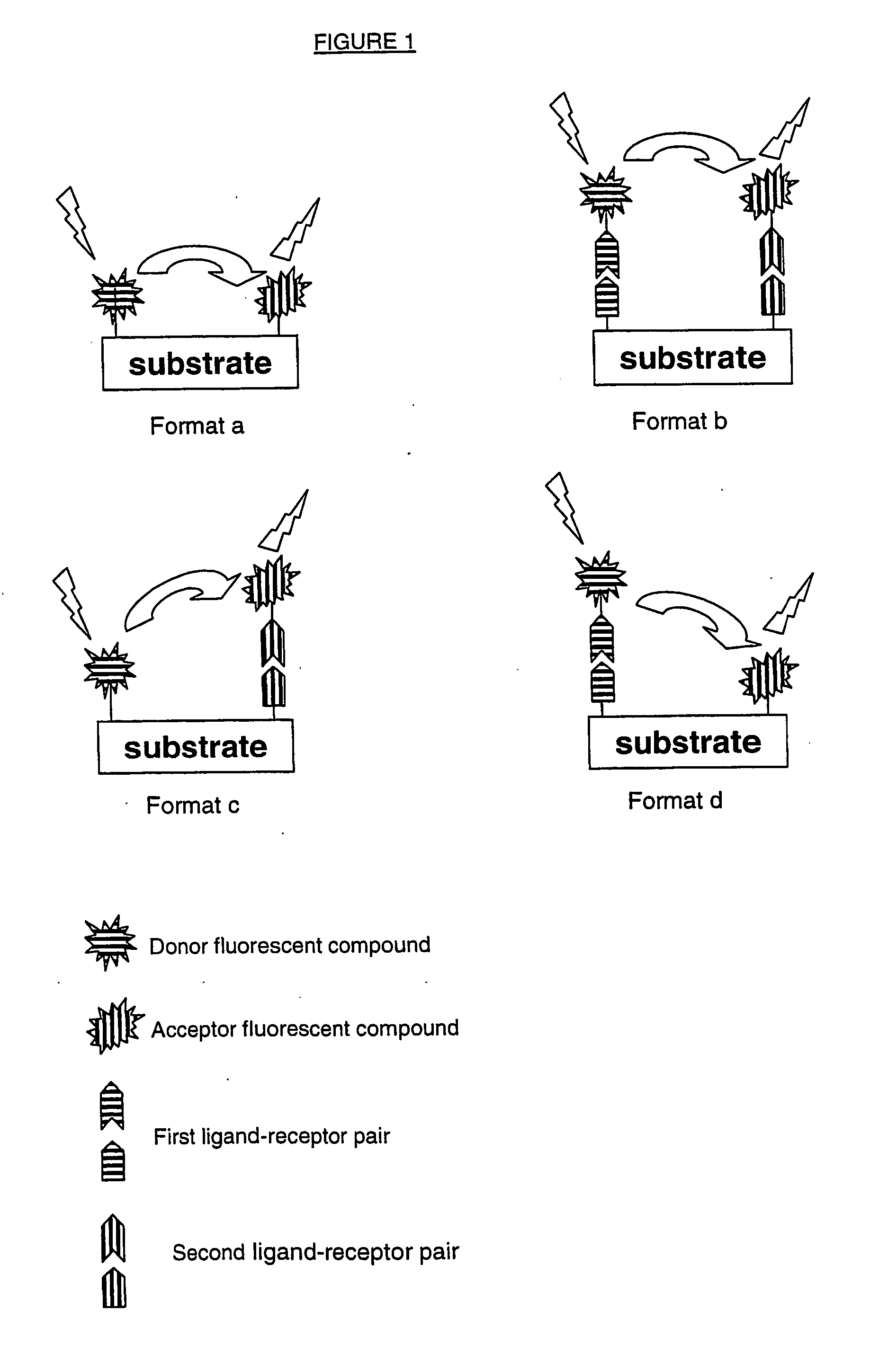
Image

Examples
example 1
Preparation of a DNP-HS-Biotin Substrate
[0104] Reagents Used: [0105] Solution of HS at 20 mg / ml: 10 mg HS+0.5 ml 10 mM PO4(Na / K) buffer, pH 7.0, 0.15M NaCl. [0106] Solution of EDC at 20 mM: 2.4 mg EDC (PIERCE)+0.625 ml 0.1 M MES buffer, pH 6.0. [0107] 47 mM 5-(biotinamide)pentylamine solution: 4.2 mg 5-(biotinamide) pentylamine (PIERCE)+0.271 ml 0.1 M MES buffer, pH 6.0. [0108] Solution of DNP-NHS at 1 mg / ml: 3 mg DNP-NHS (CIS-Bio)+1.0 ml DMSO (Sigma D8418).
[0109] Biotin Labeling [0110] 0.25 ml of a solution of HS at 20 mg / ml (Seikagaku) are mixed with 0.125 ml of 47 mM 5-(biotinamide)pentylamine solution. The mixture is incubated for 18 h at room temperature in the presence of 0.125 ml of a 20 mM EDC solution. The reaction is then stopped by adding 1.5 ml of PO4(Na / K) buffer (10 mM) containing NaCl (0.15M), pH 7. The mixture is then dialyzed for 1.5 h, using a Slide-A-Lyzer dialysis system (Pierce), against 600 ml of 0.1M PO4(Na / K) buffer, pH 7. Two further dialyses carried out f...
example 2
Determination of the Final Molar Ratios
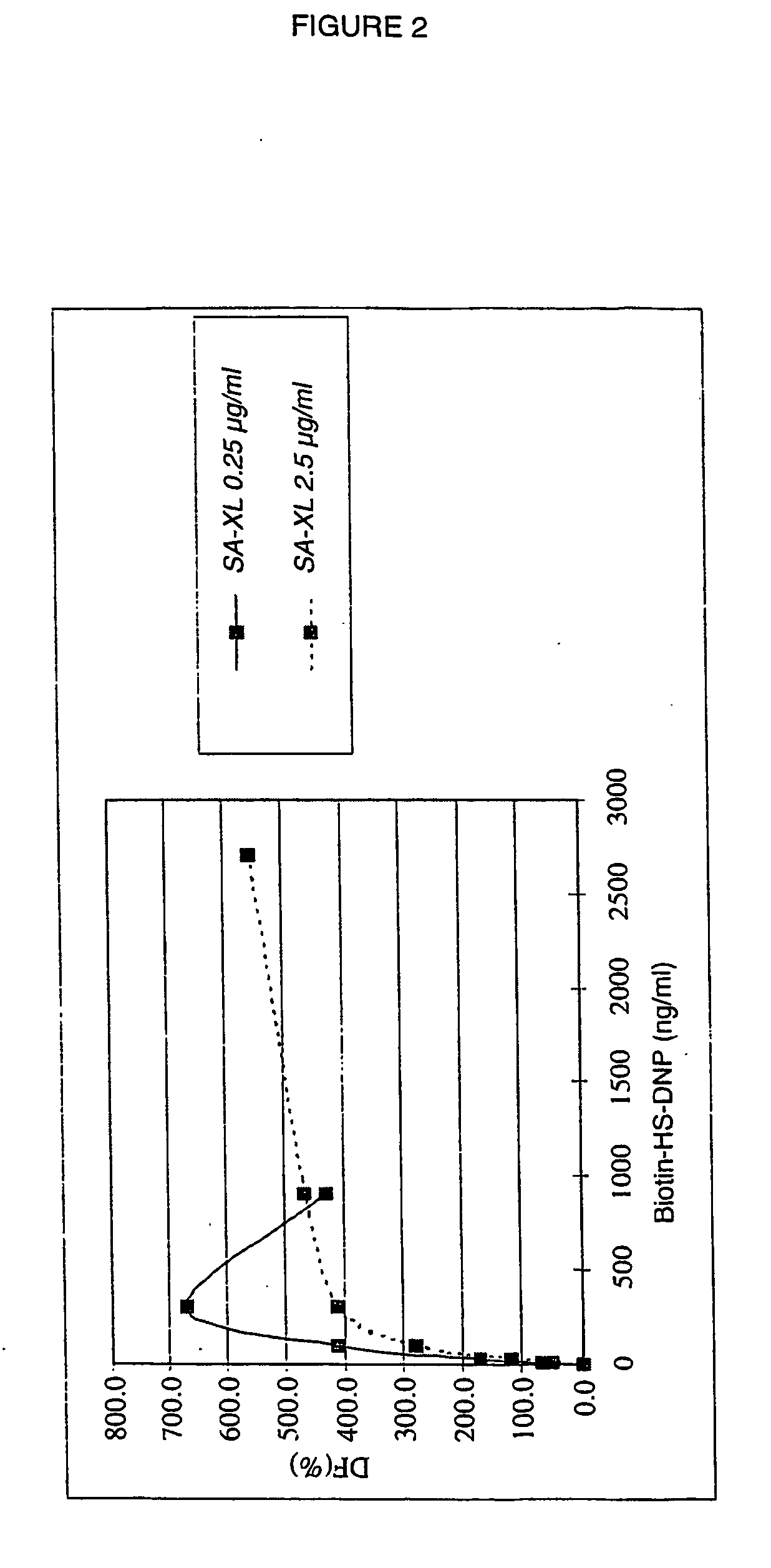
[0113] Assaying of the DNP-HS-Biotin:
[0114] A kit for assaying HSs (Byscan, Biocolor Ltd) makes it possible to determine the concentration of DNP-HS-biotin.
[0115] Assaying of the DNP:
[0116] The DNP concentration is determined by measuring the absorbance of a solution of DNP-HS-biotin at 360 nm on a spectrophotometer; the DNP concentration is determined by comparing the measured value with a standard curve.
[0117] Assaying the Biotin:
[0118] The biotin concentration is measured using an assay based on the FRET phenomenon: a first curve is established by bringing into contact known concentrations of biotin, a cryptate-streptavidin donor conjugate and an XL-biotin acceptor conjugate, and measuring the signal obtained on a Rubystar fluorimeter (BMG). A displacement curve is plotted and will be used as a standard range.
[0119] The same experiment is carried out, replacing the biotin with the DNP-HS-biotin to be assayed. The value of the signal o...
example 3
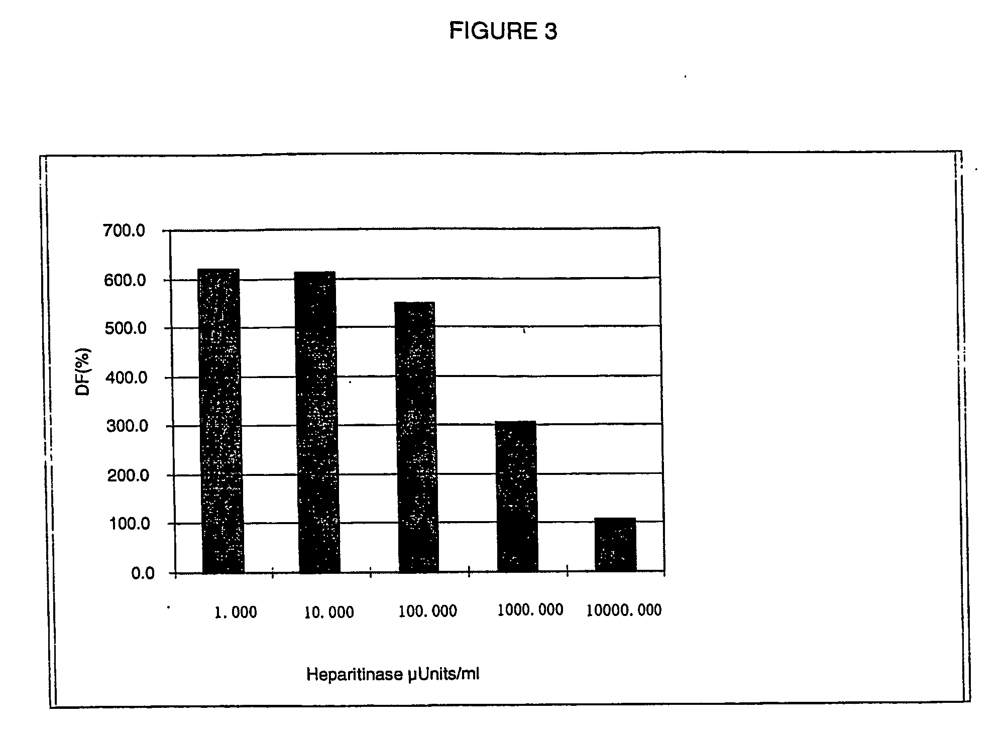
Assaying the DNP-HS-Biotin Compound Using A Homogeneous Time-Resolved Fluorescence (HTRF®) Measurement Method
[0121] The present example makes it possible to validate the use of the DNP-HS-biotin product in an assay based on the time-resolved measurement of fluorescence emitted by radiative transfer, in homogeneous medium.
[0122] Reagents Used:
[0123] Streptavidin-XL conjugate, solution at 10 μg / ml: 3.2 μl of SA-XL (CIS bio international) at 625 μg / ml+197 μl of 0.1M PO4(Na / K) buffer, pH7.0; 0.1% BSA 0.4M KF.
[0124] Streptavidin-XL conjugate, solution at 1 μg / ml: 18 μl of SA-XL (CIS bio international) at 20 g / ml+162 μl of 0.1M PO4(Na / K) buffer, pH7.0; 0.1% BSA 0.4M KF.
[0125] Anti-DNP antibody-cryptate conjugate (hereinafter referred to as aDNP-K), solution at 1 μg / ml: 4.5 μl of aDNP-K (CIS bio international) at 100 μg / ml+445 μl of 0.1M PO4(Na / K) buffer, pH 7.0, 0.1% BSA 0.4M KF.
[0126] DNP-HS-biotin solutions of varying concentration (from 22.2 to 5 400 ng / ml) are prepared from the s...
PUM
| Property | Measurement | Unit |
|---|---|---|
| Molar ratio | aaaaa | aaaaa |
| Fluorescence | aaaaa | aaaaa |
| Enzyme activity | aaaaa | aaaaa |
Abstract
Description
Claims
Application Information