

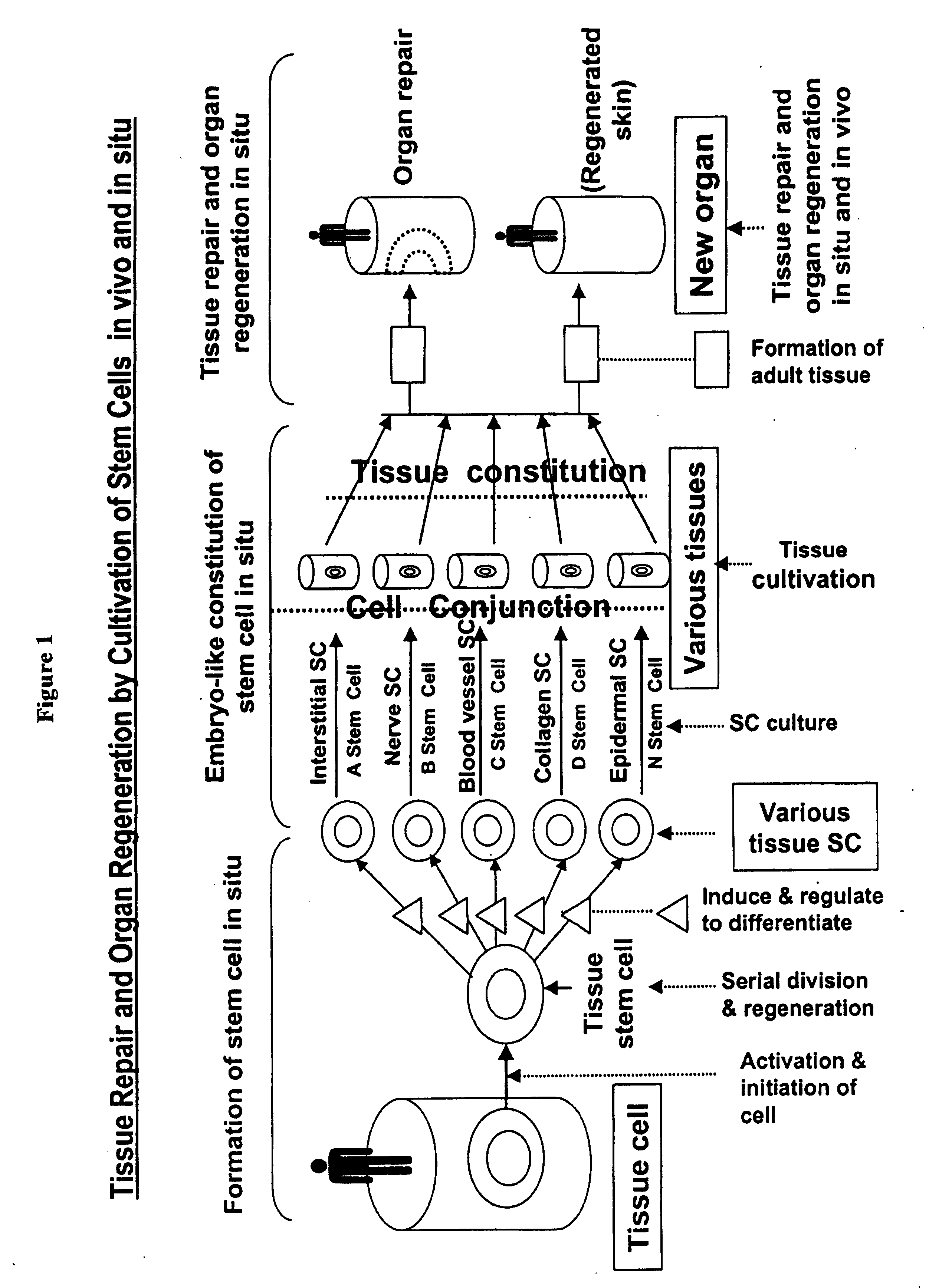
Physiological tissue repair and functional organ regeneration by cultivation of regenerative stem cells in vivo and in situ
a technology of regenerative stem cells and tissue repair, which is applied in the field of tissue engineering and organ regeneration, can solve the problems of limited time for harvesting grafts from the same site, inadequate, healthy skin donor sites are difficult to find in such patients, and the autograft method faces a few challenges and limitations, so as to reduce the viability of viable tissues
- Summary
- Abstract
- Description
- Claims
- Application Information
AI Technical Summary
Benefits of technology
Problems solved by technology
Method used
Image

Examples
example
Example 1
Manufacturing Process of an Embodiment of the Inventive Composition
[0565] The following is a description the manufacturing process of an embodiment of the inventive composition according to the present invention.
[0566] First, raw materials for the production of the composition were purified according to industrial standards. Sesame oil was filtered and transferred to an oil tank. Roots of the plant huangqin (Radix Scutellariae) were washed, cut into small pieces, pulverized, sieved, and transferred into a stock tank. Beeswax was purified by aqueous decoction.
[0567] Second, the purified sesame oil was added to a reaction tank and heated at 140-160° C. for 20 minutes with stirring, and huangqin prepared above, huangbai, and earthworm was added to the reaction tank containing sesame oil, each at a weight ratio of 10 kg:100 kg. The mixture of huangqin and sesame oil was stirred and heated at 150-160° C. for 20-30 minutes. The dreg was filtered and discarded, and the clear o...
example 2
Growth of Mammalian Cells in Culture Media Containing the Inventive Composition
[0570] In this example, in vitro experiments were designed to demonstrate that the inventive composition has unique activities in promoting proliferation and tissue-specific adhesion of normal differentiated mammalian cells and mammalian stem cells, as well as maintaining the integrity of skin structure. Skin tissue cells, hair follicle stem cells and skin pieces were sampled from rats or mice and cultured in vitro. The cell or tissue culture is divided into two groups: the control group cultured in normal cell culture media (complete MEM) and the treatment group cultured in complete MEM with the addition of the inventive composition.
[0571] An embodiment of the inventive composition, the IC prepared in Example 1, was used in the in vitro experiment.
[0572] 1) Mouse Skin Cell Culture
[0573] Mouse skin cells were harvested from fresh skin of mice immediately after sacrifice and cultured in MEM in 6-well c...
example 3
Treatment of Diabetic Skin Ulcer with the IC
[0584] Eight patients (three males and 5 females, age 40-68 yr) suffering from type II-diabetic skin ulcer were treated with the IC. Most of the patients had surface ulcer in the lower limb with ulcer areas ranging from 1% to 3% of the body surface area. The depths of ulcer in these patients either reached the dermis, hypodermnis, or the muscle layer. Necrotic tissues were surgically debrided with scissors while avoiding injury to the viable tissue surrounding the ulceric area. The IC was applied topically to the ulceric area 3-5 times a day in a sufficient amount to cover the area at about 1 mm thickness. Four patients with smaller ulceric areas healed within 1 week of the treatment; two patients with deep ulcer (which caused exposure of the tendon ligaments) healed within 2 weeks of treatment; one patient with a large surface ulcer in the buttocks (about 2% of the body surface area) healed within 3 weeks of treatment; and one patient wi...
PUM
 Abstract
Description
Claims
Application Information