Directional self-assembly of biological electrical interconnects

a biological electrical interconnect and self-assembly technology, applied in the direction of nanoinformatics, liquid/solution decomposition chemical coating, peptide sources, etc., can solve the problems of inability to nucleate microtubules in tubulin solutions, and inability to fully realize the effect of gamma-turcs and tubulin solution nucleation

Inactive Publication Date: 2007-03-15
THE ARIZONA BOARD OF REGENTS ON BEHALF OF THE UNIV OF ARIZONA
View PDF3 Cites 7 Cited by
  • Summary
  • Abstract
  • Description
  • Claims
  • Application Information

AI Technical Summary

Problems solved by technology

Such top-bottom manufacturing methods are reaching their limits in attempts to develop smaller feature sizes.
Control of interconnections emerges as one of the major challenges in the development of these bottom-up approaches.
While the process of MT growth is reasonably well understood, in vivo and in vitro MT nucleation is, however, still poorly understood.
Gamma-TuSCs can also nucleate microtubule in tubulin solutions but with lower efficiency compared with gamma-TuRCs(32).
Electroless deposition of copper onto the MT surface poses a challenge, because the commercially available plating baths contain formaldehyde which damage or destroy MTs.

Method used


Image

  • Directional self-assembly of biological electrical interconnects
  • Directional self-assembly of biological electrical interconnects
  • Directional self-assembly of biological electrical interconnects

Examples


example 1

Cloning and Expression of Tubulin

[0075] We designed and cloned a γ-tubulin fusion protein could be used to initiate microtubule growth in a precise fashion, especially after attachment to a surface of interest. Additional details of the experiments conducted can be found in Yang et al. (2006) Biotechnology Progress 22: 303-312, which is incorporated by reference herein in its entirety.

[0076] Analysis of all of the known human γ-tubulin sequences allowed the design of specific oligonucleotides (oligos) that could then be used as primers in polymerase chain reaction (PCR) to clone γ-tubulin. In addition, those oligos that were designed to be used to PCR amplify the γ-tubulin would also provide the recombinant form with sequences that allow it to be made as a recombinant fusion with glutathione —S— transferase (GST) sequence tag. Specifically, the human sequence that was used to design primers was from NCBI (NM—001070, Homo sapiens tubulin, γ-1, TUBG1). They are:

5′-GGAATTCTGCCGAGGG...

example 2

Surface Functionalization

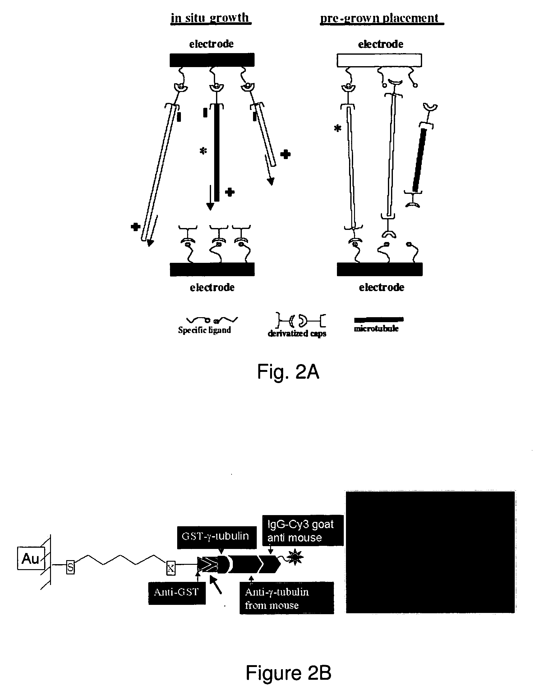
[0089] A protein nucleation method for MT growth from gold substrates has been developed based on the self-assembly of reactive alkanethiols together with the engineered fusion protein. Oxidized silicon wafers were patterned with gold electrodes, followed by treatment with piranha solution to clean organic contaminants and to activate the gold surface. An anti-GST antibody was bound to a SAM of carboxylic acid-terminated alkanethiols on the gold surface through the carboxylic acid group at the end of the alkyl chains (FIG. 2B). Anti-GST is a specific antibody for selective binding of GST attached to recombinant proteins, in our case, the microtubule-nucleating fusion protein, GST-γ-tubulin. Additionally, a specific antibody that binds γ-tubulin is then recognized by a secondary antibody that has a fluorescent tag (IgG-Cy3) and is used to quantify the extent of the formation of the protein assembly. Strong fluorescence from a functionalized gold surface on a...

example 3

MT Growth

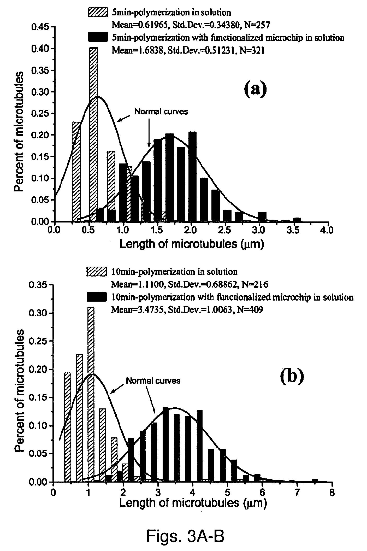
[0091] For this study, we used tubulin (>99% pure) prepared from bovine brain extracts and modified with covalently linked fluorescein (Cytoskeleton Inc). The fluorescein-modified tubulin was stored at −70° C. in storage buffer (pH 6.8; 80 mM piperazine-N,N′-bis 2-ethanesulfonic acid sequisodium salt (PIPES), 1 mM magnesium chloride (MgCl2); 1 mM ethylene glycol-bis(b-amino-ethyl ether) N,N,N′,N′-tetra-acetic acid (EGTA) and 1 mM guanosine 5′-triphosphate (GTP)).

[0092] In-vitro MT assembly was performed in PEM 80 buffer (80 mM PIPES, 1 mM EGTA, 4 mM Magnesium chloride (MgCl2), using KOH to adjust PH to 6.9) using a final concentration of tubulin at 0.25 mg / ml (2.3×10⁶ M). Polymerization was initiated by the addition of GTP (final concentration is 0.25 mM) in the presence of taxol (final concentration is 10 μM). Taxol reduces the competing “depolymerization” process.

[0093] In order to test the specificity of the interaction between MTs and the γ-tubulin functionalized sub...


PUM

PropertyMeasurementUnit
inner diameteraaaaaaaaaa
pHaaaaaaaaaa

Abstract

Microtubules are excellent candidates for the fabrication of nanostructures, including nanowires. A method for controlled nucleation and growth of microtubules on substrates (e.g., gold on a silicon wafer) is provided. The substrate is functionalized with a nucleating agent for microtubule growth. The method can be employed to generate nanoscale structures on substrates or between substrates by additional attachment of MT capture agents which function to capture the ends of growing MT to form connecting MT structures. The method can be used to form 2- and 3-D structures on or between substrates and can function to establish interconnects between nanoscale devices or molecular electronic devices and electrodes. A specific method for metallization of biological macromolecules and structures in provides which can be beneficially applied to metallized the MT formed by the growth and capture method. The metallization method is biologically benign and is particularly useful for copper metallization of MTs.

Description

CROSS REFERENCE TO RELATED APPLICATIONS [0001] This application claims the benefit of U.S. provisional patent application Ser. No. 60 / 677,734, filed May 4, 2005, the entire contents of which are incorporated by reference herein.STATEMENT REGARDING U.S. GOVERNMENT FUNDING [0002] This invention was funded by the United States Government under National Science Foundation contract No. 0303863. The U.S. government has certain rights in this invention.BACKGROUND OF THE INVENTION [0003] In the semiconductor industry there has been an ongoing trend towards smaller and smaller electronic circuits and devices. This technology has been sustained by modifying the capabilities of manufacturing processes such as photolithography. Such top-bottom manufacturing methods are reaching their limits in attempts to develop smaller feature sizes. Implementation of a bottom-up method is now sought after for the manufacture of nanoscale electronic circuits. A bottom-up method utilizes the self-assembly natu...

Claims


Application Information

Patent Timeline
Patent Type & Authority Applications(United States)
IPC IPC(8): C12Q1/68H01L21/00
CPCB81B2207/07B82B3/00B82Y5/00B82Y10/00B82Y30/00B82Y40/00H01L2221/1094C23C18/1844C23C18/2086C23C18/40C23C18/44H01L21/76838C07K14/47
Inventor DEYMIER, PIERREJONGEWAARD, IAN N.HOYING, JAMES B.GUZMAN, ROBERTORAGHAVAN, SRINI
Owner THE ARIZONA BOARD OF REGENTS ON BEHALF OF THE UNIV OF ARIZONA
Why Eureka
Try Eureka