Methods and Devices for Quantitative Viral Assays

a quantitative and viral assay technology, applied in the field of methods and microfluidic devices, can solve the problems of difficult repeatability, inability to precisely control the flow environment, and inability to accurately and accurately measure the viral replication ability, and achieve the effect of extending the spread and better reflecting the replication ability of the virus

Active Publication Date: 2007-05-17
WISCONSIN ALUMNI RES FOUND
View PDF1 Cites 3 Cited by
  • Summary
  • Abstract
  • Description
  • Claims
  • Application Information

AI Technical Summary

Benefits of technology

[0007] The present invention takes advantage of the discovery that the extent of spread of viral progeny from single infected cells is greater during comet formation than during plaque formation, and that measurement of viral infectivity based on comet formation is faster and more sensitive. Comet size also better reflects virus replicating ability than plaque size. This is because, if a sufficiently high flow exits, each virus particle in the virus ‘plume’ released by the initial infected cell could in principle find a susceptible host cell, near or far from the virus source, and eventually create an expanse of dead, dying, or detached cells corresponding with the magnitude of the plume (the number of progeny released from the initial infection). By contrast, the plume from an initially infected cell in a traditional plaque assay is confined to the distance to which the virus particles can diffuse. For example, diffusivity in water for VSV particles is about 2×10⁻⁸ cm2 / sec (Ware et al., 1973, J. Virol.: 11: 141-145), so over a typical 6-hour VSV infection cycle, the diffusional spread for the viral particles in agar is less than about 0.02 cm, corresponding to an area of 10−3 cm2, which contains an estimated 300 cells. On the other hand, an initial infected cell typically releases 1000 to 5000 viral progeny, most of which thus will not find an uninfected cell over the time scale of a secondary infection cycle. In short, virus particles originating from the initial infected cell have a better chance in the comet assay, compared to the standard plaque assay, of initiating a secondary infection that will be detected, be quantified, and contribute to further spread. In addition, because the “comets” are more easily observable, a comet based assay is faster than the classical plaque based assays.

Problems solved by technology

While such convection or evaporation-driven flows in standard six-well culture dishes are simple to implement, they lack precise control over the flow environment, and precise, repeatable and quantitative measurements are difficult.

Method used


Image

  • Methods and Devices for Quantitative Viral Assays
  • Methods and Devices for Quantitative Viral Assays
  • Methods and Devices for Quantitative Viral Assays

Examples


example 1

Comet Assay Measures Drug Susceptibility with High Sensitivity

1. Methods

[0066] Cell culture. Baby hamster kidney (BHK-21) cells were originally obtained from Dr. I. Novella (Medical College of Ohio) and grown as monolayers at 37° C. in a humidified atmosphere containing 5% CO2. Growth medium consisted of Minimum Essential Medium Eagle with Earle's Salts (MEM, Cellgro), 10% fetal bovine serum (FBS, Hyclone), and 2 mM Glutamax I (Glu, Gibco). Cells were subcultured approximately every fourth day. For subculture, monolayers were rinsed with Hank's Balanced Salt Solution (HBSS, Hyclone), incubated in 0.05% trypsin / 0.53 mM EDTA (Gibco) for 5 minutes, and replated in fresh growth medium at a 1:30 dilution. No antibiotics were used and cells were subcultured no more than 100 passages to minimize the artifacts due to cell senescence. Viability of cell populations, as determined by trypan blue exclusion, at the time of experiments always approached 100%. In all the antiviral assays used i...

example 2

Cells Grow in Microchannels

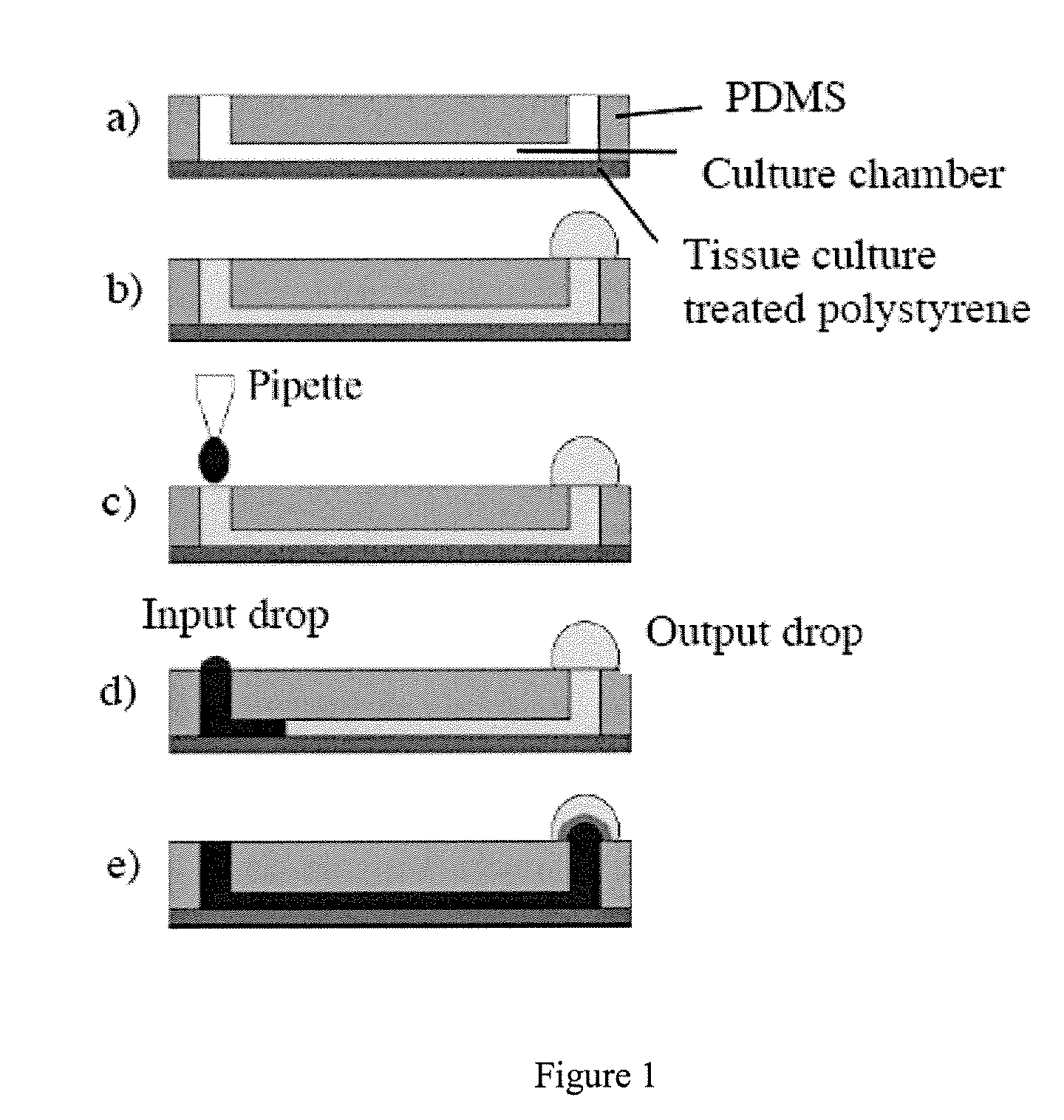
[0075] Microchannels were made from poly(dimethylsiloxane) (PDMS), with a configuration of two ports joined by a channel, as shown in FIG. 1. Typical total volume of the channel and two ports is 8 microliters. The ‘floor’ of the microchannel is typically the bottom of a culture well in a standard six-well polystyrene culture plate. Cells added to the channel through one port adhere to the ‘floor’ and eventually flatten out and form a monolayer.

[0076] Baby hamster kidney (BHK21) cells were seeded into microchannels at 1000 cells / mm2, in the presence of growth medium. Phase contrast images were obtained at indicated times (hours post cell loading=HPCL=number of hours after cells were loaded into the microchannel). Representative images are shown in FIG. 7.

[0077] Quantification of cell growth in microchannels. A BHK cell suspension was injected into a channel through one port. Such cell loading was performed with several channels in parallel. At indicated ...

example 3

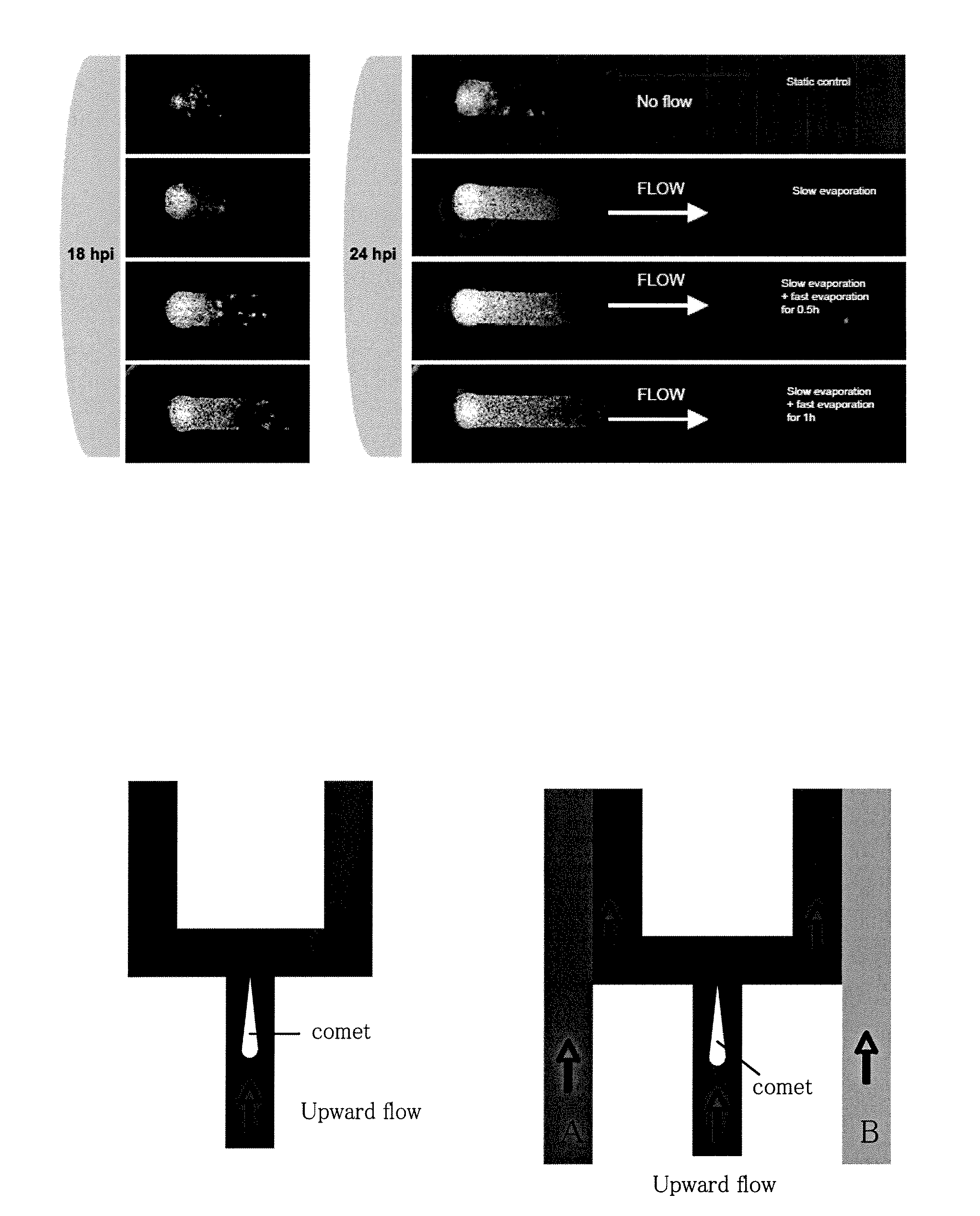
Flow-Enhanced Spread of Virus Infections in Microchannels

[0081] BHK cells were loaded into the microchannels at 4·106 cells / ml to create confluent monolayers. Control and test channels were all inoculated with one microliter of virus inoculum containing fewer than 10 plaque forming units; this one microliter volume is sufficient to displace the initial volume of fluid in the port. Both ports of the control “no-flow” sample were covered with pieces of PDMS to minimize evaporation from the channel and thereby maintain a static fluid environment. The test samples were inoculated in the same manner, but flows were induced by creating imbalances in the evaporation rates from the two ports, as follows. A 5-microliter droplet of 2% medium was added on one port. Therefore, when evaporation occurred, medium flowed from the droplet through the channel to the other port. To achieve slow-flow conditions samples were placed in a ‘high-humidity’ (or slow evaporation) incubator, containing a pan ...


PUM

PropertyMeasurementUnit
flow velocityaaaaaaaaaa
volumeaaaaaaaaaa
volumeaaaaaaaaaa

Abstract

A method for quantifying infectious particles of a virus in a sample comprises providing a layer of host cells of the virus, contacting the layer of host cells with a preparation of the sample, and culturing the cells under conditions wherein the cells are submerged in a thin layer of liquid culture medium, and wherein the virus infects host cells and releases its progeny from said infected host cells, imposing a flow of the liquid medium, wherein the spread of the viral progeny to uninfected host cells is enhanced, culturing the cells under conditions to allow further virus infection and viral gene expression, wherein infected host cells develop an observable indication of viral gene expression, and determining the number of infected host cells, whereby the number of infectious particles of the virus in the sample is quantified. The method may be used for measuring viral growth rate or for screening for antiviral compounds. Also provided are microfluidic devices suitable for the inventive method.

Description

FIELD OF THE INVENTION [0001] This invention relates to method and microfluidic devices for assaying viral growth, infectivity, and resistance to anti-viral substances, and for screening for antiviral compound agents. BACKGROUND OF THE INVENTION [0002] For many diagnostic and / or treatment purposes, it is highly desirable to be able to measure the number of infectious viral particles in a sample or preparation. For example, rapid and accurate measurement of viral growth is necessary for treatment of viral infections and for monitoring whether a therapeutic strategy is effective in inhibiting viral growth, or whether the virus has developed resistance to compounds used for treatment. Also, as viral vectors are primary delivery vehicles for gene therapy, it is critical that the quantity of the viral particles or dosage in a clinical preparation be rapidly and accurately determined. [0003] Although total particle measurement can be made by techniques such as electron microscopy of viral...

Claims


Application Information

Patent Timeline
Patent Type & Authority Applications(United States)
IPC IPC(8): C12N7/00C12N5/00C12N7/01C12N5/02
CPCC12N7/00C12N2760/20211C12N15/86C12N2760/16051C12N2770/36251C12N2795/14151C12Q1/00C12Q1/70
Inventor YIN, JOHNSHU, YING
Owner WISCONSIN ALUMNI RES FOUND
Why Eureka
Try Eureka