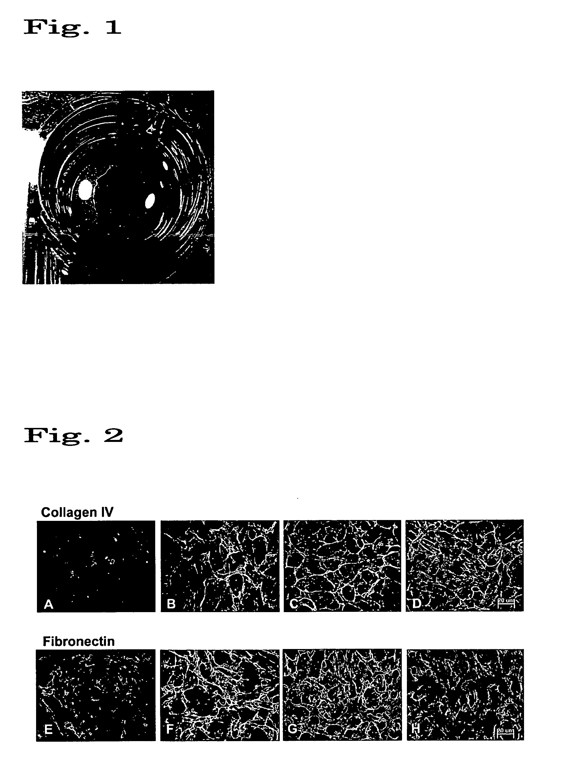
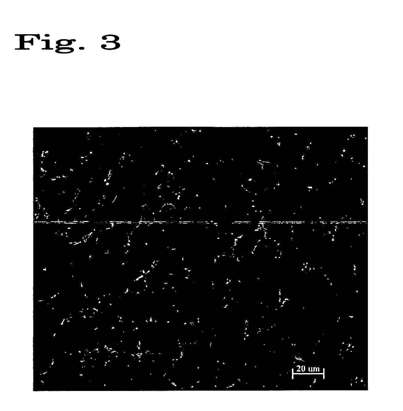
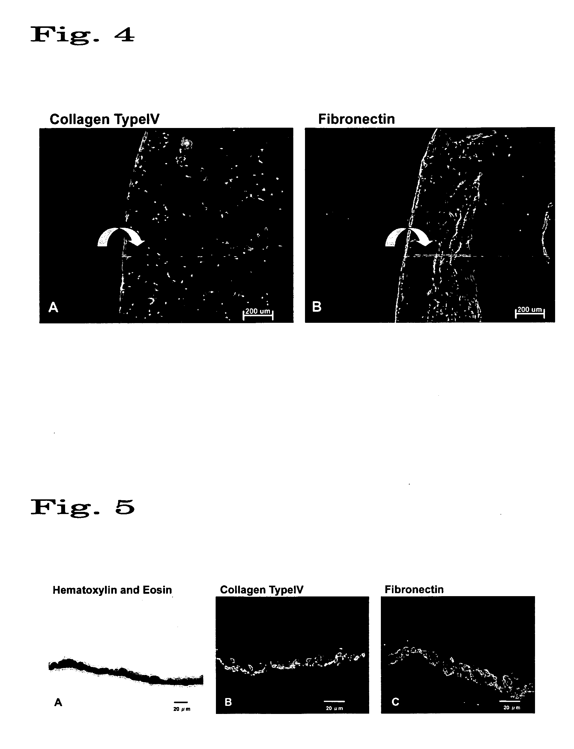
Regenerated corneal endothelial cell sheets, processes for producing the same, and methods of using the same

a corneal endothelial cell and cell sheet technology, applied in the field of regenerated corneal endothelial cell sheets, can solve the problems of corneal tissue swelling, corneal tissue swelling, and the number of donors in japan is still considerably smaller

Active Publication Date: 2007-06-28
CELLSEED +2
View PDF16 Cites 26 Cited by
  • Summary
  • Abstract
  • Description
  • Claims
  • Application Information

AI Technical Summary

Benefits of technology

[0010] In order to attain the stated objects, the present inventors engaged in R&D activities taking various angles of study. As a result, the inventors found that a regenerated corneal endothelial cell sheet that would adhere very well to a living tissue could be obtained by a process comprising the steps of cultivating corneal endothelial cells under specified conditions on a cell culture support comprising a substrate having its surface covered with a specif

Problems solved by technology

If the remaining corneal endothelial cells are too few, the pumping function of the corneal endothelial tissue is no longer adequate and the corneal tissue swells to develop diseases such as corneal endothelial disorder and bullous keratopathy.
However, the number of donors in Japan is still considerably smaller than that of patients and while there are annually about 20,000 patients who need keratoplasty, only a tenth of them (ca.
Although keratoplasty is a virtually established procedure, it suffers the problem of shortage in donors, giving rise to the need for the development of a next-generation medical procedure.
However, it has been pointed out that the recovery

Method used


Image

  • Regenerated corneal endothelial cell sheets, processes for producing the same, and methods of using the same
  • Regenerated corneal endothelial cell sheets, processes for producing the same, and methods of using the same
  • Regenerated corneal endothelial cell sheets, processes for producing the same, and methods of using the same

Examples


examples

[0054] On the following pages, the present invention is described in greater detail by reference to examples which are by no means intended to limit the scope of the invention.

examples 1 and 2

[0055] To a commercial 3.5 cmφ cell culture dish (FALCON 3001 manufactured by Becton Dickinson Labware), a coating solution having N-isopropylacrylamide monomer dissolved in isopropyl alcohol to give a concentration of 40 wt % (Example 1) or 45 wt % (Example 2) was applied in a volume of 0.1 ml. By applying electron beams with an intensity of 0.25 MGy, an N-isopropylacrylamide polymer (PIPAAm) was immobilized on the surface of the culture dish. After the irradiation, the culture dish was washed with ion-exchanged water to remove the residual monomer and the PIPAAm that did not bind to the culture dish; the culture dish was then dried in a clean bench and sterilized with an ethylene oxide gas to provide a cell culture support material. The coverage of PIPAAm was found to be 1.6 μg / cm2 (Example 1) or 1.8 μg / cm2 (Example 2).

[0056] In a separate step, a corneal endothelial tissue was collected from the corneal limbus of a white rabbit under deep anesthesia by the usual method and the c...

example 3

[0060] A cell culture support material was prepared as in Example 1. Subsequently, a coating solution having an acrylamide monomer containing N,N-methylene bisacrylamide (1 wt % / acrylamide monomer) dissolved in isopropyl alcohol to give a concentration of 5 wt % was applied over the support material in a volume of 0.1 ml. A metallic mask having a diameter of 1.8 cm was superposed and while being kept in that state, the support material was exposed to electron beams with an intensity of 0.25 MGy, whereupon an acrylamide polymer (PAAm) was immobilized except in the area under the metallic mask. After the irradiation, the culture dish was washed with ion-exchanged water to remove the residual monomer and the PAAm that did not bind to the culture dish; the culture dish was then dried in a clean bench and sterilized with an ethylene oxide gas to provide a cell culture support material.

[0061] In the next step, by repeating the same procedure as Example 1, a corneal endothelial tissue was...


PUM


Abstract

An improved process for producing a regenerated corneal endothelial cell sheet, comprising the steps of allowing corneal endothelial cells collected from a tissue to be cultivated on a cell culture support having its surface covered with a polymer of which the hydrating force varies in a temperature range of 0-80° C., and after the culture,
(1) adjusting the temperature of the culture solution to the temperature at which the polymer on the substrate surface is hydrated,
(2) bringing the cultured corneal endothelial cell sheet into close contact with a carrier, and
(3) detaching the sheet together with the carrier.
The regenerated corneal endothelial cell sheet obtained by the process will adhere very well to living tissues.

Description

TECHNICAL FIELD [0001] This invention relates to regenerated corneal endothelial cell sheets in biology, medicine and other fields, as well as processes for producing such sheets, and therapeutic methods using them. BACKGROUND ART [0002] The corneal tissue consists of five layers, a corneal epithelium layer on the outermost surface, Bowman's membrane, a corneal stromal layer, Descemet's membrane, and a corneal endothelium layer. The innermost corneal endothelium layer serves as a liner on the corneal tissue, and Descemet's membrane corresponds to the basement membrane of the corneal endothelial layer which is composed of adhesive proteins. The function of corneal endothelial cells is to pump water out of the corneal stroma into the anterior chamber counteracting the swelling pressure of the stroma, presumably due to the pumping action as assisted by Na—K ATPase. Human corneal endothelial cells will not usually undergo cell division in the living body, and denatured or exfoliated end...

Claims


Application Information

Patent Timeline
IPC IPC(8): A61K35/30C12N5/08A61L27/00A61L27/38A61L27/50C12N5/071
CPCA61L27/3808A61L27/3839A61L27/3895A61L27/50A61L2430/16C12N5/0621C12N2539/10A61L27/38A61L27/44
Inventor OKANO, TERUONISHIDA, KOHJIYAMATO, MASAYUKI
Owner CELLSEED
Why Eureka
Try Eureka