Method to determine 3-d elemental composition and structure of biological and organic materials via atom probe microscopy

a technology of atom probe and organic material, which is applied in the direction of separation process, filtration separation, instruments, etc., can solve the problems of requiring massive calculations, requiring large calculations, and reducing the accuracy of the results, so as to increase the microscope field of view and accelerate the analysis time

Inactive Publication Date: 2007-08-09
IMAGO SCI INSTR
View PDF3 Cites 18 Cited by
  • Summary
  • Abstract
  • Description
  • Claims
  • Application Information

AI Technical Summary

Benefits of technology

[0023] This invention is made practical by the development of improved 3-D atom probe microscopes, as exemplified by Imago Scientific Instruments' local electrode atom probe (LEAP). Such improvements include substantial increases in the microscope field of view, the geometry of the specimens that may be examined, and much more rapid analysis times. Nonetheless, the invention described herein can be applied to conventional atom probes by those skilled in the art Thus, the subject invention also pertains to determining the structure of biological and organic materials with other types of atom probes and related instruments.

Problems solved by technology

Each of these methods has certain inherent limitations, especially when applied to biologically important molecules:
Computational Modeling: The major difficulty for computationally determining 3-D structures based upon knowledge of the primary amino acid sequence of a protein is that these require massive calculations.
Such an assumption may not be warranted.
Finally, computational methods for determining protein structure from amino acid sequences cannot be generally extended to determine the conformation of other biomolecules such as nucleotides, nucleoproteins, carbohydrates, glycosaminoglycans, and proteoglycans.
Nor can models designed to predict protein 3-D structure be extended to predict the 3-D structure of non-biological organic materials such as synthetic polymers.
However, obtaining such information is a very slow and laborious process.
It can take months to convert X-ray crystallographic data into a corresponding 3-D structure because diffraction patterns are extremely complex (e.g., as many as 25,000 diffraction spots from a single small protein).
Additional difficulties in crystallographic analysis arise because the molecule under study must first be crystallized into a highly regular crystal with millimeter or near-millimeter dimensions.
The single act of preparing a suitable crystal to begin the analysis can itself take months, years, or even prove impossible.
Further still, the resolution obtained is limited by the quality of the crystal analyzed.
Due to the time required for crystallization and the collection and analysis of the diffraction data, determining the structure of a single, relatively simple macromolecule can easily take a year or more.
Hence the molecule of interest must be isolated from its normal environment thereby potentially altering its conformation.
The higher resolution method of liquid state NMR (in contrast to solid state NMR) is generally limited to molecules of a molecular weight no larger than about 40 kD due to the requirement that the molecules rapidly tumble in solution.
High-resolution NMR is not able to analyze high-molecular weight (MW) biomolecules, such as many larger proteins and macromolecular complexes.
Although solid-state NMR does not have the size and solubilization limitations of liquid-state NMR, the lower resolution limitation of solid-state NMR is only able to provide nanometer-level resolution, and not the necessary angstrom-level resolution.
Finally, the analysis of NMR spectra is complex, time-consuming, and less than straightforward.
High-Resolution Microscopy: Current scanning and transmission electron microscope technology is not able to provide the necessary sub-nanometer resolution with biological and organic materials.
While high-resolution transmission electron microscopies (e.g., TEM, intermediate- and high-voltage transmission electron microscopy [IVEM and HVEM], scanning transmission electron microscopy [STEM], and scanning electron microscopies [SEM]) can provide instrumental resolution in the angstrom range with some robust materials (e.g., metals and ceramics), this is not possible with organic materials because low atomic number organic elements provide low intrinsic contrast and are easily destroyed by electron beams.
This is 10-to 50-fold poorer resolution than the required for atomiclevel resolution.
However, angstrom-level resolution has not been achieved for biological materials using any type of electron microscopy.
Finally, electron microscopy does not directly provide compositional or elemental information.
With carbon and the other low atomic number elements found in biological and other organic materials, the obtainable spatial resolution for elemental mapping is rarely better than about 10 nanometers and provides limited sensitivity.
Scanning probe microscopies, including atomic force microscopy (AFM), scanning tunneling micrscopy (STM), near-field scanning optical microscopy (NSOM), and related methods, are also not suitable for providing 3-D atomic resolution and elemental mapping of biological and organic materials.
They are not capable of gathering information on the layers underlying the surface.
Scanning probe microscopies also do not provide information on elemental composition.
Planar structures like wafer-processed materials, e.g., microelectronic materials, are often difficult to cut into atom probe specimens because the structures of interest exist only in a very thin layer on the surface of the specimen, a specimen that is often less than about 10 micrometers (microns) thick.
The sample preparation techniques described in U.S. Pat. Nos. 6,576,900 and 6,700,121, however, are not suitable for analyzing biological materials, organic materials, and otherwise non-conductive materials.

Method used


Image

  • Method to determine 3-d elemental composition and structure of biological and organic materials via atom probe microscopy
  • Method to determine 3-d elemental composition and structure of biological and organic materials via atom probe microscopy
  • Method to determine 3-d elemental composition and structure of biological and organic materials via atom probe microscopy

Examples


Embodiment Construction

(1) Overview:

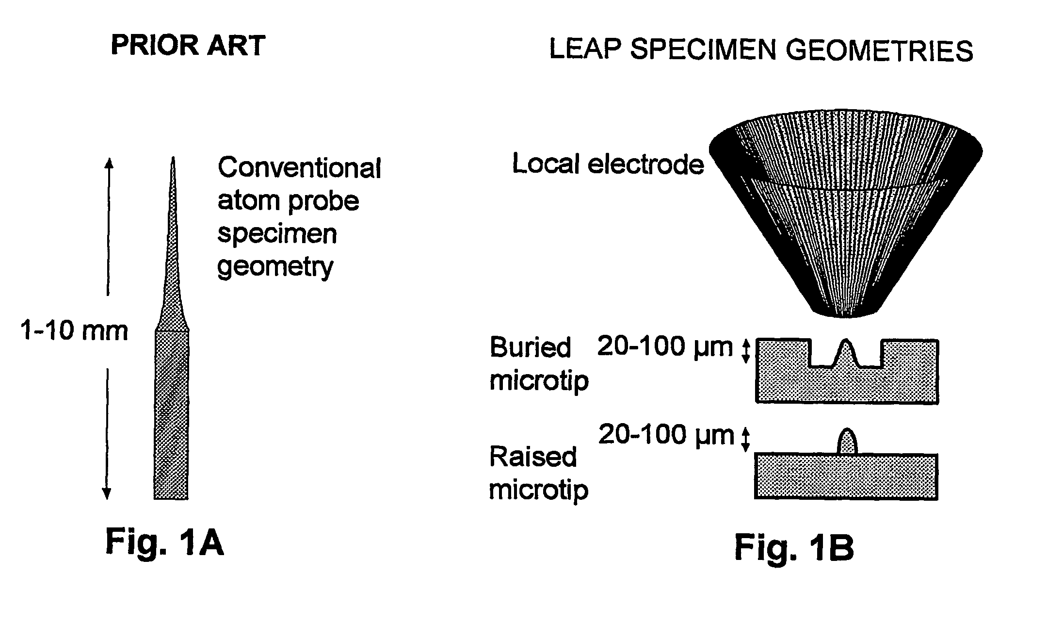
[0063] The vast majority of specimens previously analyzed using atom probes have been metals. The reasons for this are two-fold: 1) the need for the specimen to have high electrical conductivity; and 2) the need for the specimen to have sufficient physical strength to be shaped into a millimeter-long, needle-shaped specimen (the required geometry). Consequently, there is only a modest history of atom probe analysis of biological or organic specimens. Field ion microscopy (FIM) images of freeze-dried tRNA dimers adsorbed onto iridium tips were reported by Machlin in 1975 (Machlin, Freilich et al. 1975), while Panitz has provided what appears to be the most recent report of FIM and field desorption imaging, which was applied to ferritin (Panitz 1981; Panitz 1982) and unstained DNA (Panitz 1983). Other than a few additional publications by these research groups, this is essentially the entire history of biological atom probe and FIM imaging.

[0064] A major problem discus...


PUM

PropertyMeasurementUnit
thickaaaaaaaaaa
diameteraaaaaaaaaa
thicknessaaaaaaaaaa

Abstract

Disclosed are methods to prepare a specimen for microanalysis, the specimens so prepared, and methods to analyze the specimens by atom probe microscopy. The specimens are prepared by embedding a specimen within an electrically conductive matrix to yield an embedded specimen; and then forming regions on the embedded specimen into shapes suitable for microanalysis by an atom probe.

Description

CROSS-REFERENCE TO RELATED APPLICATIONS [0001] Priority is hereby claimed to provisional application Ser. No. 60 / 492,789, filed Aug. 6, 2003, the entire content of which is incorporated herein.FIELD OF THE INVENTION [0002] The present invention relates to methods and instrumentation to determine the three-dimensional structure and elemental composition of biological materials at the molecular, near-atomic, and atomic level using three-dimensional atom probe microscopy and related methods. BACKGROUND [0003] In the manufacture of many modem devices containing microscopically thin layers of different materials, and / or zones of different materials segregated on a microscopic scale, it is important to be able to study the different layers and / or zones with analytical equipment after the deposition. As examples, it is often useful to be able to microscopically analyze the structures of semiconductor microelectronic devices; magnetic thin film memory storage devices (such as read / write har...

Claims


Application Information

Patent Timeline
Patent Type & Authority Applications(United States)
IPC IPC(8): G01N33/48B81B1/00
CPCB81B1/008B82Y15/00G01Q60/00B82Y35/00B82Y30/00G01N1/00G01N1/36H01J37/285
Inventor GOODMAN, STEVEN L.KELLY, THOMAS F.KREMER, JOHN J.
Owner IMAGO SCI INSTR
Why Eureka
Try Eureka