Ocular therapeutic agent delivery devices and methods for making and using such devices

a technology of ocular therapeutic agent and delivery device, which is applied in the field of controlled release of ocular implant device, can solve the problems of side effects, negligible actual absorption of drug in ocular tissues, and failure of oral therapies for the eye to provide sustained release of drug into the eye, etc., and achieves the effects of high release rate or loading dose, high release rate, and constant drug delivery ra

Inactive Publication Date: 2007-08-16
GOVEMMENT OF THE US REPRESENTED BY THE SEC DEPT OF HEALTH & HUMAN
View PDF53 Cites 31 Cited by
  • Summary
  • Abstract
  • Description
  • Claims
  • Application Information

AI Technical Summary

Benefits of technology

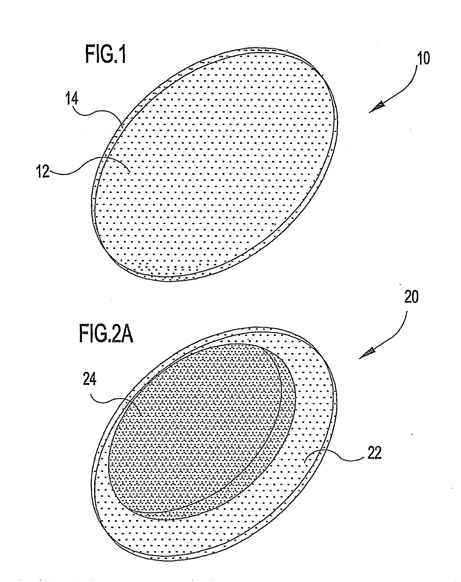
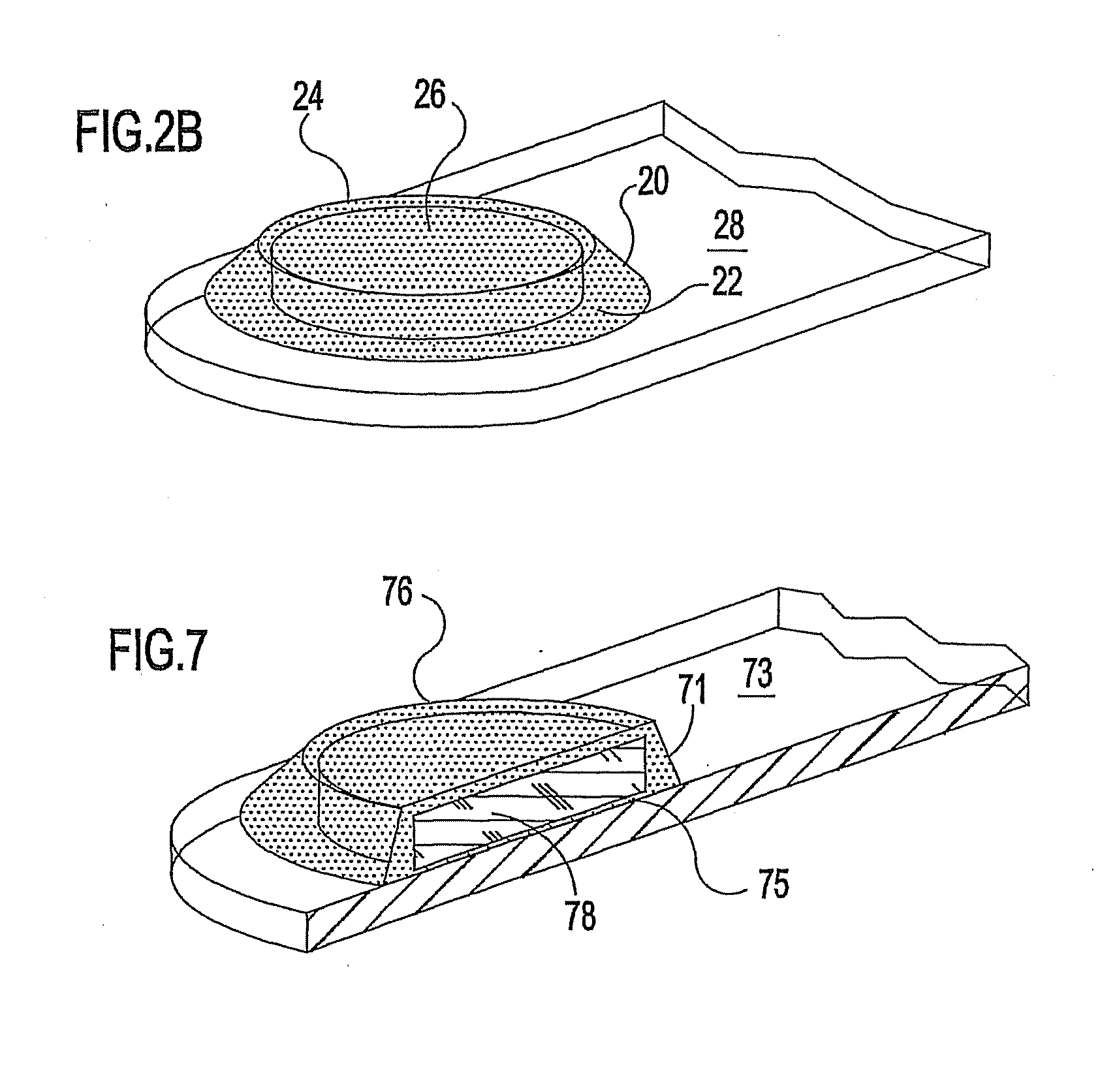
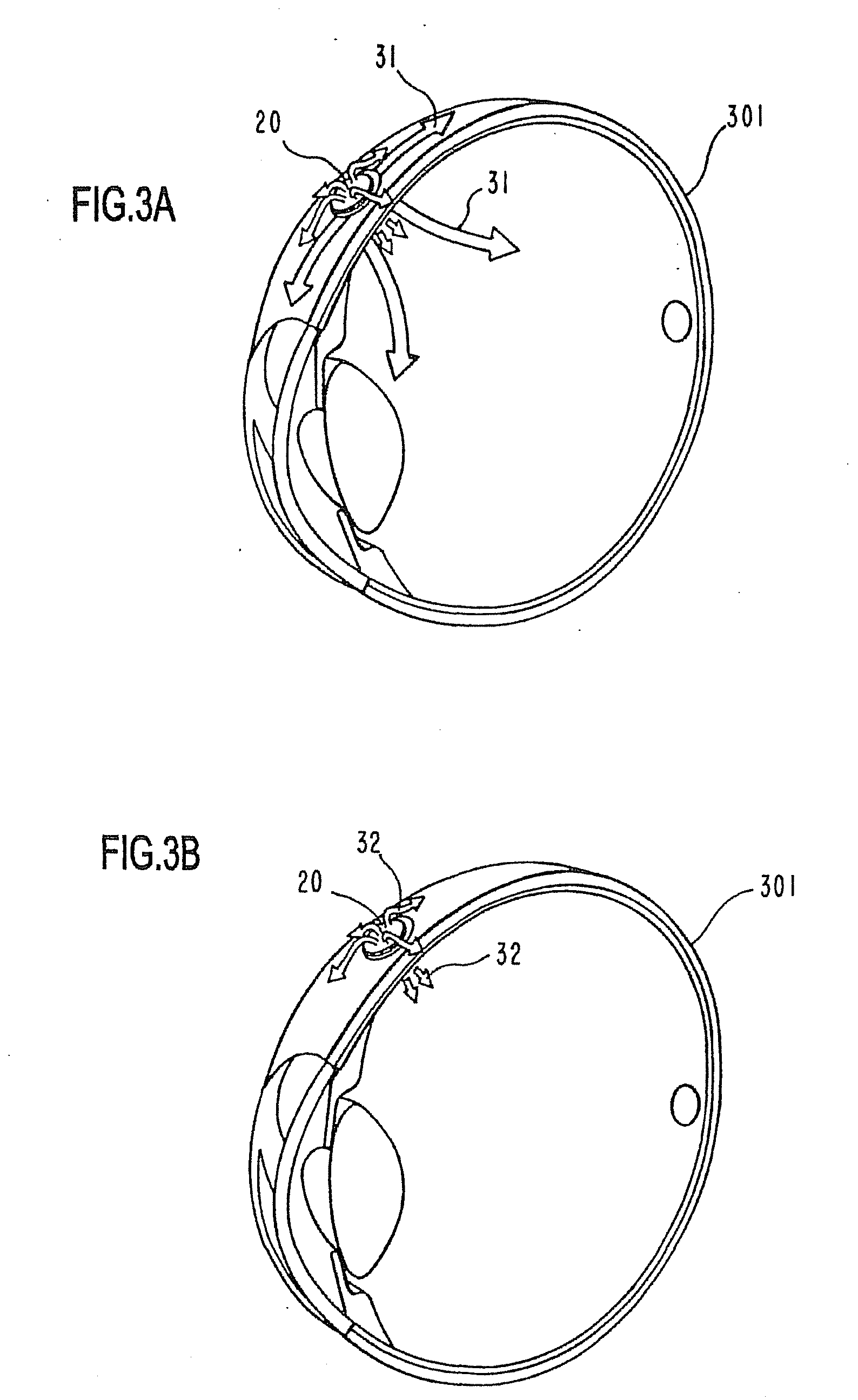
[0047] The implant subassembly of the intraocular reservoir implants of the invention provide a sustained, substantially constant delivery rate of drug over a prolonged period. The intraocular reservoir implants also can be modified to form dual mode release devices. For instance, in a dual mode configuration, additional therapeutic agent could be dispersed in the silicone fluid before being used to encapsulate the drug core to create an initial higher release rate, or loading dose; alternatively, additional amounts of the drug could be dispersed in or attached as a discrete inlay member onto a separate silicone adhesive used to attach a surface of the reservoir implant subassembly to a suture stub or the like. Alternatively, multi-drug therapy could be provided by including a drug different from the drug core in the silicone surrounding the pellet or, in or on the silicone adhesive used to affix the implant reservoir subassembly, each comprising an encapsulated drug core, can be attached to a common suture stub to provide concurrent delivery of different drugs or additive introduction of a common drug.
[0048] As another dual mode embodiment of the reservoir impla

Problems solved by technology

For instance, oral therapies for the eye fail to provide sustained-release of the drug into the eye.
Instead, oral therapies often only result in negligible actual absorption of the drug in the ocular tissues due to low bioavailability of the drug.
Moreover, adverse side effects have been associated with systemic administration of certain drugs to the eyes.
For instance, systemic treatments of the eye using the immune response modifier cyclosporine A (CsA) have the potential to cause nephrotoxicity or increase the risk of opportunistic infections, among other concerns.
However, therapeutic levels of the drug are not achieved and sustained in the middle or back portions of the eye.
This is a major drawback, as the back (posterior) chamber of the eye is a frequent site of inflammation or otherwise the site of action where, ideally, ocular drug therapy should be targeted for many indications.
Although only about ten percent of people with AMD have the wet form, it poses a much greater threat to vision.
With the wet form of the disease, rapidly growing abnormal blood vessels known as choroidal neovascular membranes (CNVM) develop beneath the macula, leaking fluid and blood that destroy light sensing cells and causing a blinding scar tissue, with resultant sever loss of central vision.
However, in that the macula is located at the back of the eye, treatmen

Method used


Image

  • Ocular therapeutic agent delivery devices and methods for making and using such devices
  • Ocular therapeutic agent delivery devices and methods for making and using such devices
  • Ocular therapeutic agent delivery devices and methods for making and using such devices

Examples


example 1

[0208] This example illustrates the preparation of a matrix, implant of the invention useful for subconjunctival implants.

[0209] 4.5 g of superhydrolyzed polyvinyl alcohol (Airvol 125, Air Products and Chemicals, Inc., Allentown, Pa., U.S.A.) was added to 30 ml of molecular biology grade water in an assay tube that was then tightly closed. The tightly closed assay tube was placed in a beaker of boiling water until the density becomes uniform (generally about 3-7 hrs). Since the assay tube was tightly closed, the contents could not evaporate. Water was periodically replaced in the beaker to keep the water height near the height of water in the assay tube. The assay tube was centrifuged for 1 minute at 1000-4000 rpm to degass the mixture. This formed a 15 wt % solution of superhydrolyzed polyvinyl alcohol.

[0210] Separate premixtures were prepared using each of cyclosporine A and 2ME2 as the therapeutic agent. Each therapeutic agent was separately premixed in a solution of hydroxypro...

example 2

[0213] This example illustrates the preparation of another matrix implant of the invention, which is useful as an intravitreal implant. Alternatively, this matrix implant can be used for an inlay used in combination with reservoir implants of the invention described elsewhere herein.

[0214] Preparation of 50% Superhydrolyzed PVA, 6% 2ME2, 0.05% HPMC Matrix Implant:

[0215] The polymer drug mixture was prepared in a 3 cc syringe (the tip sealed with a Luer lok and a HPLC septum). The plunger was removed. A drug emulsion was prepared by adding 63.8 mg 2ME2 and 0.5 mg hydroxypropyl methylcellulose (E4M, Dow Chemical) to 2 ml molecular grade biological water. The emulsion was mixed with a magnetic stirrer over night, and then it was added to 1 g superhydrolyzed PVA (Airvol 125; Air Products and Chemicals, Inc., Allentown, Pa., U.S.A.) in the syringe. The mixture was stirred until uniform and then placed into a water bath at about 100° C. for 60 minutes. The sample with the syringe was th...

example 3

[0216] A 1×1×2 mm matrix implant was prepared using poly(ethylene vinyl) acetate (EVA) in place of superhydrolyzed PVA in the subconjunctival implant.

[0217] Preparation of 30% EVA, 6% 2ME2, 0.05% HPMC Matrix Implants:

[0218] The polymer drug mixture was prepared in a 3 cc syringe (the tip sealed with a Luer lok and a HPLC septum). The plunger is removed. A drug emulsion is prepared by adding 38.3 mg 2ME2 and 0.3 mg HPMC (E4M, Dow Chemical) to 2 ml methylene chloride. The emulsion is mixed over night with a magnetic stirrer and then transferred to a 10 ml vial containing 0.6 g EVA (Elvax 40W, Dupont). The mixture was stirred with a magnetic stirrer until it becomes too viscous. The magnetic stirrer was then removed and the mixture was left overnight. The sample was centrifuged as needed to degas the specimen. The specimen was poured onto a glass plate and permitted to dry for 48 hours under vacuum. The implants were then cut to size using a razor blade, e.g., multiple 1×1×2 mm slabs...


PUM

PropertyMeasurementUnit
Timeaaaaaaaaaa
Timeaaaaaaaaaa
Thicknessaaaaaaaaaa

Abstract

Ocular implant devices (10, 20, 121) for the delivery of a therapeutic agent to an eye (101, 301) in a controlled and sustained manner. Dual mode and single mode drug delivery devices (10, 20, 121) are illustrated and described. Implants (10, 20) suitable for subconjunctival placement are described. Implants (121, 10, 20) suitable for intravitreal placement also are described. The invention also includes fabrication and implementation techniques associated with the unique ocular implant devices (10, 20, 121) that are presented herein.

Description

CROSS-REFERENCE TO RELATED APPLICATIONS [0001] This patent application is a continuation of copending U.S. patent application Ser. No. 10 / 471,468, filed Sep. 12, 2003 [pending], which is a PCT national phase application of PCT / US0207836 filed Mar. 14, 2002, which is a continuation-in-part of U.S. patent application Ser. No. 09 / 808,149, filed Mar. 15, 2001 [issued Mar. 30, 2004 as U.S. Pat. No. 6,713,081].FIELD OF THE INVENTION [0002] The present invention generally relates to local therapies for the eye and, more particularly, to controlled-release ocular implant devices, including methods for making and using such devices, for delivery of therapeutic agents to the eye. BACKGROUND OF THE INVENTION [0003] In the treatment of many diseases and disorders of the eye, and especially in the case of degenerative or persistent conditions, implantable sustained-release delivery devices have been desired that would continuously administer a therapeutic agent to the eye for a prolonged period ...

Claims


Application Information

Patent Timeline
IPC IPC(8): A61F2/02A61F9/00A61F9/007A61K9/00
CPCA61F9/0017Y10S425/808A61K9/0051A61F9/00781A61P27/02A61K9/5026A61K9/5031A61K9/5047A61K9/5089A61K31/565A61K31/58A61K38/13B29D11/023B29K2083/00B29K2827/18
Inventor ROBINSON, MICHAEL R.CSAKY, KARL G.NUSSENBLATT, ROBERT B.SMITH, JANINE A.YUAN, PENGSUNG, CYNTHIAFRONHEISER, MATTHEW P.KIM, HYUNCHEOL
Owner GOVEMMENT OF THE US REPRESENTED BY THE SEC DEPT OF HEALTH & HUMAN
Why Eureka
Try Eureka