Preparation method of biotinylated protein and detection method using the same

Inactive Publication Date: 2007-08-16
CELLFREE SCI
View PDF0 Cites 6 Cited by
  • Summary
  • Abstract
  • Description
  • Claims
  • Application Information

AI Technical Summary

Benefits of technology

[0042] The method for preparing a biotinylated protein according to the present invention enables a rapid analysis of polyspecimen, because it does not require a step of removing free biotin derivatives.

Problems solved by technology

According to these methods, however, in a detection step, one or more washing operations are required, thus these methods have defects in sample-processing capacity, and the like.
Because of this, in various detection methods, particularly in detection steps of ALPHA, the free biotin-removing step before detection=makes the operations of whole assay troublesome, and, as a result, they had problems in rapidly analyzing polyspecimen.

Method used


Image

  • Preparation method of biotinylated protein and detection method using the same

Examples


example 1

Preparation of Wheat Embryo Extract

(1) Preparation of Wheat Embryo

[0115] Chihoku wheat seeds of Hokkaido origin, or Chikugo-izumi seeds of Ehime Prefecture origin were added to a mill (supplied by Fritsch Co.: Rotor Speed Millpulverisette type 14) at a rate of 100 g per minute, and the seeds were gently pulverized at 8,000 rpm. After a fraction containing embryo with germinability was recovered with a sieve (mesh size: from 0.7 to 1.00 mm), a floating fraction containing the embryo with germinability was recovered by flotation using a mixture of carbon tetrachloride and cyclohexane (a volume ratio of carbon tetrachloride:cyclohexane=2.4:1), and the organic solvent was removed by drying at room temperature and then contaminating impurities such as seed coat were removed by air blasting at room temperature to give a crude embryo fraction.

[0116] Subsequently, using a belt-type color separator BLM-300K (manufacturer: Anzai Manufacturing Co., Ltd., selling agency: Anzai Sogyo), embr...

example 2

Preparation of Biotinylating Enzyme

[0123] pEU vector to which a base sequence coding a biotinylating enzyme from E. coli genome was inserted was prepared. Subsequently, a transcription template was prepared using the pEU vector as a template and using PCR. The transcription template was added to a transcription reaction solution [final concentrations: 80 mM of HEPES-KOH (pH 7.8), 16 mM of magnesium acetate, 10 mM of dithiothreitol, 2 mM of spermidine, 2.5 mM of 4NTPs (4 kinds of nucleotide triphosphates), 0.8 U / μl of RNase inhibitor, and 1.6 U / μl of SP6 RNA polymerase], and the transcription was conducted at 37° C. for 3 hours (Proc Natl Acad Sci USA, 2002, vol 99, pl4652-14657: Sawasaki, T et al.). All of the obtained mRNA pellets were added to the wheat embryo extract (200 O.D.) obtained in the above-mentioned Example 1, and the protein synthesis was conducted at 26° C. for 15 to 20 hours.

[0124] It was confirmed by using a radio isotope ([14C]-Leu) that the above biotinylating ...

example 3

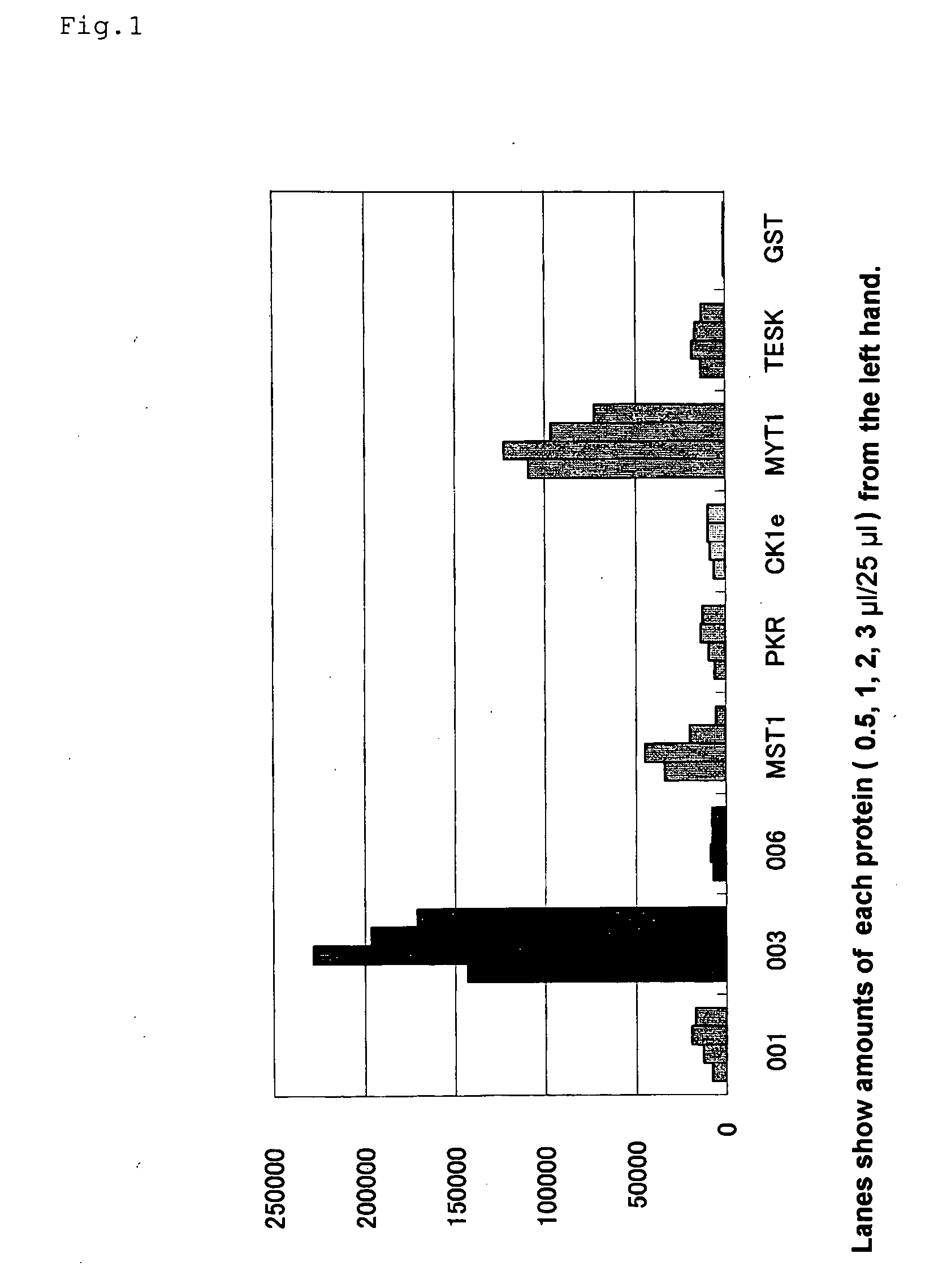
Preparation of a Translation Template of Biotinylated Protein (001-006)

[0125] In relation to the translation template mRNA, a vector which was a biotinylated protein transcription template in which a gene represented as a gene number of 001-006 was fused with a biotin tag (pEU-biotinylated tag-001-006) was prepared. Based on the vector, a PCR product containing Ω sequence part of tobacco mosaic virus (TMV) was used as a template. The transcription template was added to a transcription reaction solution [final concentrations: 80 mM of HEPES-KOH (pH 7.8), 16 mM of magnesium acetate, 10 mM of dithiothreitol, 2 mM of spermidine, 2.5 mM of 4NTPs (4 kinds of nucleotide triphosphates), 0.8 U / μl of RNase inhibitor, 1.6 U / μl of SP6 RNA polymerase], and the reaction was conducted at 37° C. for 3 hours. The obtained RNA was extracted with phenol / chloroform, precipitated with ethanol, and purified with Nick Column (supplied by Amersham Pharmacia Biotech Inc.) to give a translation template.

[...


PUM

PropertyMeasurementUnit
Timeaaaaaaaaaa

Abstract

The present invention presents construction of a detection method requiring no step of removing free biotin during preparation of a biotinylated protein having a biotin tag, in a detection method of a substance interacting with a protein, and studied various preparation methods of the biotinylated protein. In order to solve the above-mentioned problem, the present inventor has found that in a cell-free protein synthesizing system, in particular, a wheat embryo cell-free protein synthesizing system, when biotinylation is performed during or after protein's synthesis, the biotinylation of the protein can be attained in an remarkably lower concentration of the biotin than that in the conventional biotinylation operations, and has accomplished the present invention by using the protein having the biotin tag in each detection system.

Description

BACKGROUND OF THE INVENTION [0001] 1. Field of the Invention [0002] The present invention relates to a preparation method of a biotinylated protein and to a method for detecting a substance interacting with a biotinylated protein, using the protein. More particularly, the invention relates to a detection method wherein a biotinylated protein is prepared in a cell-free protein synthesizing system, and a substance interacting with the protein is detected. [0003] This application claims the priority of Japanese Patent Application Nos. 2006-182785 and 2005-377840, which are incorporated herein by reference. [0004] 2. Related Background of the Invention [0005] A variety of methods are utilized for analyzing a biomolecule interaction in an intracellular reaction. Such methods include, for example, ELISA (Enzyme Linked Immunosorbent Assay), DELFIA (Dissociation Enhanced Lanthanide Fluoroimmunoassay), and SPA (Scintillation Proximity Analysis). These methods are referred to as so-called “he...

Claims


Application Information

Patent Timeline
IPC IPC(8): G01N33/53C07K14/47
CPCC07K1/13G01N33/532G01N2500/00G01N33/58G01N33/543
Inventor ENDO, YAETASAWASAKI, TATSUYAMATSUBARA, YUKO
Owner CELLFREE SCI
Why Eureka
Try Eureka