
Methods for modulating glutamate receptors for treating neuropsychiatric disorders comprising the use of modulators of serum and glucocorticoid inducible kinases

a glutamate receptor and neuropsychiatric disorder technology, applied in the direction of drug compositions, anti-noxious agents, extracellular fluid disorders, etc., can solve the problems of elusive signaling pathway from pl3-k to ampa receptor abundance in the cell membrane, and many problems remain to be solved, so as to enhance the abundance of ampa subunit glur1 protein and increase glutamate-induced currents

Inactive Publication Date: 2007-08-16
MERCK PATENT GMBH
View PDF5 Cites 4 Cited by
  • Summary
  • Abstract
  • Description
  • Claims
  • Application Information

AI Technical Summary

Benefits of technology

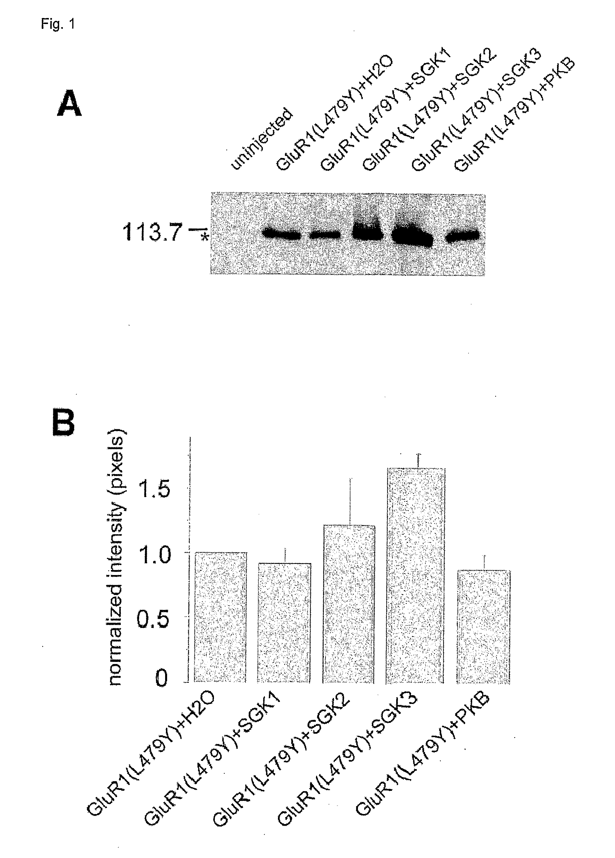
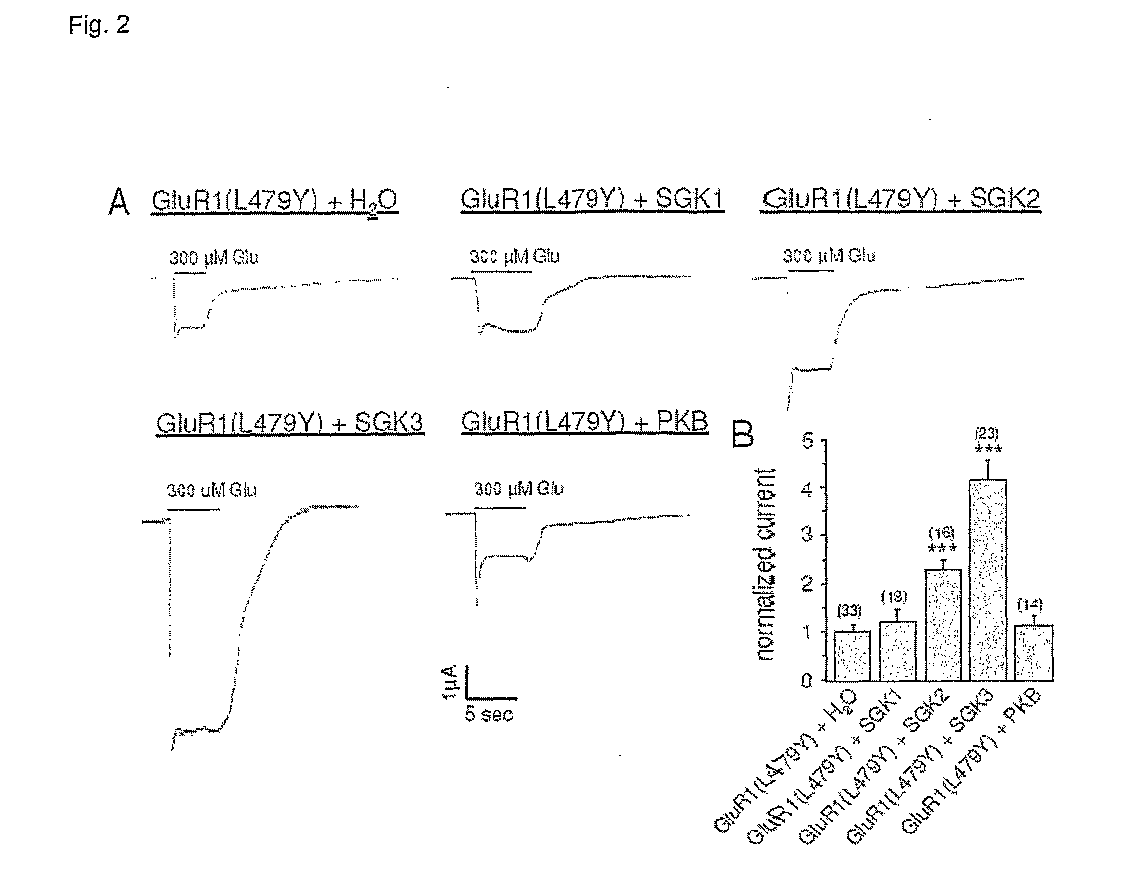
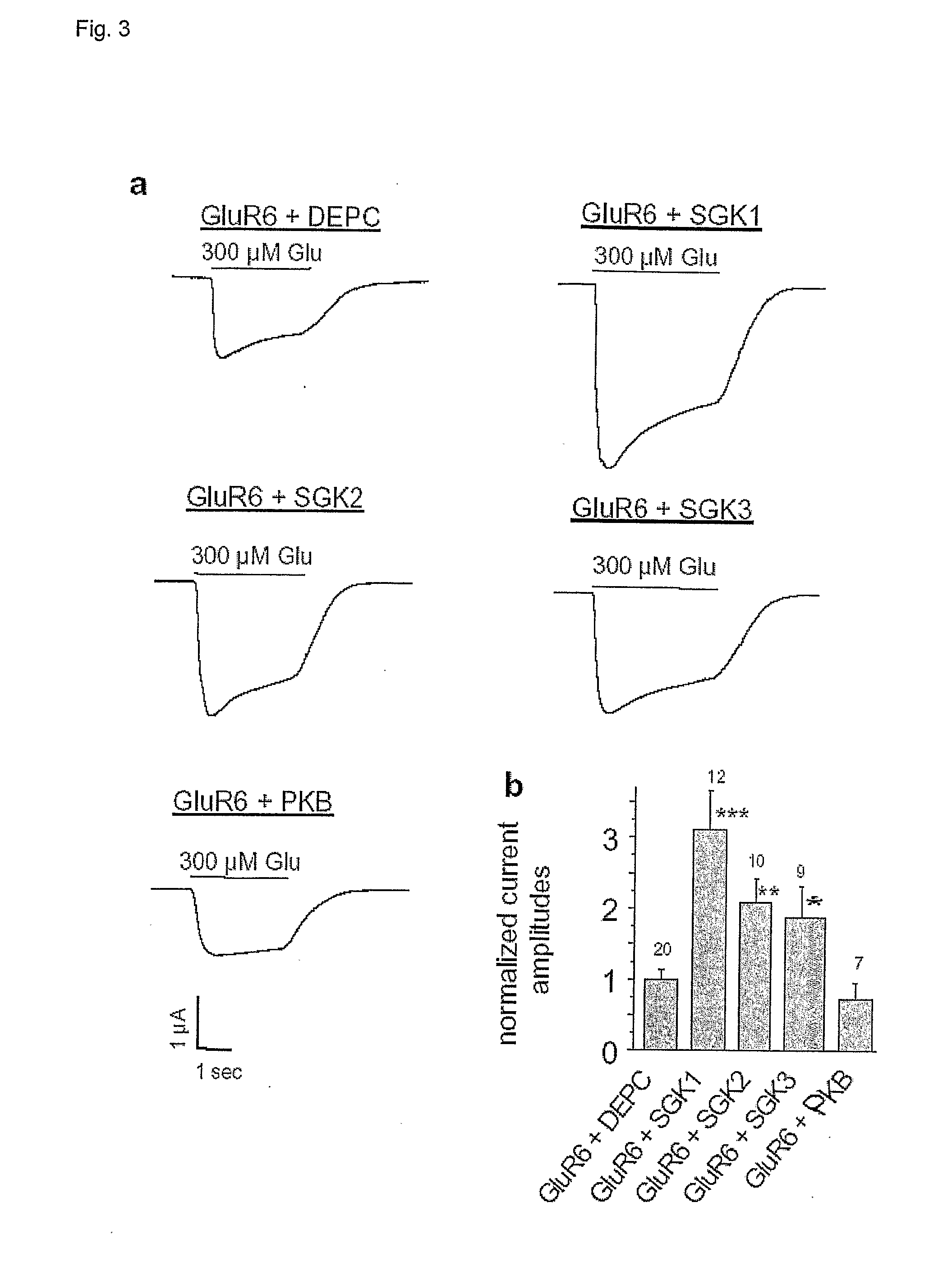
[0012] While SGK3 is a powerful regulator of GluR1, SKG1 is involved in GluR6 activation. SGK3 enhances the abundance of GluR1 in the plasma membrane and increases GluR1-mediated glutamate-induced currents.
[0016] It is furthermore shown that to a lesser extent SGK2 but not SGK1 increases glutamate-induced currents by enhancing the abundance of the AMPA subunit GluR1 protein in the cell membrane of Xenopus oocytes expressing rat GluR1.
[0026] The present observations revealed a novel mechanism in the regulation of the GluR1 subunit of AMPA receptors. The delivery of GluR1 to the neuronal surface is regulated by activation of NMDA receptors, leading to Ca2+ entry (M. Sheng, M. J. Kim, Science 298, 776, 2002) with subsequent activation of Pl3-kinase (M. S. Perkinton, J. K. Ip, G. L. Wood, A. J. Crossthwaite, R. J. Williams, J. Neurochem. 80, 239, 2002). Activation of Pl3-kinase triggers a signaling cascade eventually leading to activation of SGK3, which then enhances the protein abundance of GluR1 in the cell membrane. SGK3 leads to a stabilized GluR1 in the membrane thus preventing its retrieval and subsequent degradation and / or enhances trafficking of protein to the cell membrane. Therefore the present observations describes for the first time that SGK2 and SGK3 substantially contributes to the fine tuning of GluR1 abundance.
[0034] The inventive regulatory mechanism involving the new identified kinases is a powerful regulator of GluR6. SGK1 enhances the abundance of GluR6 in the plasma membrane and increases GluR6 mediated glutamate-induced currents. Thus, SGK1 participates in the regulation of kainate receptor trafficking, synaptic plasticity and neuronal excitability.

Problems solved by technology

However, many issues remain to be solved in order to understand the mechanism driving neuropsychiatric diseases.
However, the signaling pathway from Pl3-K to AMPA receptor abundance in the cell membrane remained elusive.

Method used


Image

  • Methods for modulating glutamate receptors for treating neuropsychiatric disorders 
comprising the use of modulators of serum and glucocorticoid inducible kinases
  • Methods for modulating glutamate receptors for treating neuropsychiatric disorders 
comprising the use of modulators of serum and glucocorticoid inducible kinases
  • Methods for modulating glutamate receptors for treating neuropsychiatric disorders 
comprising the use of modulators of serum and glucocorticoid inducible kinases

Examples


example 1

Electrophysiological Measurements in Xenopus Oocytes

[0048] Oocytes of stages V-VI were surgically removed from the ovaries of Xenopus laevis as described elsewhere (Seebohm, Sanguinetti, Pusch, 2003). Oocytes were injected with either 4 ng of GluR1 or GluR3 or GluR6 cRNA or with or without 6 ng SGK1 or SGK2 or SGK3 or PKB cRNA using a Nanoliter 2000 injector (WPI inc., Florida, USA). Standard two-electrode voltage clamp recordings were performed 5-8 days after cRNA injection with a TurboTec 03 amplifier (npi, Tamm, Germany) and an interface DIGIDATA 1322A from Axon Instruments. Data analyses were done with pClamp 9.0 / clampfit 9.0 software (Axon inc.), and Origin 6.0 software (Microcal). Agonist solutions were prepared in ND-96 buffer (in mM, NaCl, 96; CaCl2, 1,8; KCl, 2,0; MgCl2, 1,0 and HEPES-NaOH, 5, pH 7.2 with NaOH). Current and voltage electrodes were filled with 3 M KCl and had resistances of 0.5-1.5 MΩ. Oocytes were held at −70 mV and agonist (300 μM glutamate) was applied b...

example 2

Labeling of Cell Surface Proteins Using Biotinylated ConA

[0049] To identify the fraction of receptor protein inserted in the plasma membrane, surface proteins were tagged with biotinylated ConA and isolated by streptavidin / sepharose-mediated precipitation of the biotinyl-ConA / protein complex. Briefly, intact oocytes were incubated in 10 μM biotinyl-ConA (Sigma, München, Germany) for 30 min at room temperature. After five 10-min washes in normal frog Ringer, intact oocytes were homogenized with a Teflon pestle in H-buffer (20 μl / oocyte; 100 mM NaCl, 20 mM Tris-HCl, pH 7.4, 1% Triton X-100, 1 mM phenylmethylsulphonyl fluoride plus a mixture of proteinase inhibitors (Complete™ tablets, Boehringer)) and were kept at 4° C. for 1 h on a rotator. After centrifugation for 60 s at 16000×g, the supernatants were supplemented with 20 μl washed streptavidin-sepharose beads (Sigma, München, Germany) and incubated at 4° C. for 3 h on the rotator. The streptavidin-sepharose beads were then pellet...

example 3

Gel Electrophoresis and Western Blotting

[0050] Proteins from homogenized oocytes were separated by SDS electrophoresis and transferred to nitrocellulose filters. Blots were blocked in 1× PBS containing 5% milk powder for at least 1 hour at room temperature. For the detection of GluR1, GluR3 or GluR6, primary rabbit immunoaffinity purified GluR1, GluR3 or GluR6 antibody (1 μg / μl, Upstate) and secondary horseradish peroxidase-conjugated donkey anti-rabbit antibody (1:1000 dilution, Amersham Bioscience) were used. For verification of protein leves, Ponceau red staining was performed.


PUM

PropertyMeasurementUnit
apparent molecular weightaaaaaaaaaa
flow rateaaaaaaaaaa

Abstract

Modulation of the activity of serum and glucocorticoid inducible kinases to restore glutamate receptor activity. Also disclosed are methods and compounds useful for the detection and treatment of neuropsychiatric disorders.

Description

FIELD OF THE INVENTION [0001] A method for altering glutamate receptor activity comprising, contacting cells expressing serum and glucocorticoid inducible kinases SGK1, SGK2 or SGK3 with a substance that modulates glucocorticoid inducible kinases. Furthermore the invention relates to the diagnosis and treatment of diseases related to glutamate receptor up- or down-regulation. BACKGROUND OF THE INVENTION [0002] Neurons continually modify the relative expression, function, and subcellular localization of neurotransmitter receptors to maintain and fine-tune neurotransmission. Among the excitatory receptor systems modified are members of the AMPA family of the ionotropic glutamate receptor (GluR) that include subunits GluR1 thru GluR4. AMPA receptors are involved in a variety of diseases as epilepsy, Alzheimer's disease, Parkinson's disease, and Rasmussen's encephalitis. Rasmussen's encephalitis is a progressive disorder that is characterized by severe epilepsy, hemiplegia, dementia and...

Claims


Application Information

Patent Timeline
Patent Type & Authority Applications(United States)
IPC IPC(8): A61K31/573A61K31/165A61K31/519C12N5/07C12N5/079
CPCA61K31/519A61K31/165A61P25/00A61P25/08A61P25/18A61P25/22A61P25/24A61P25/28A61P25/30A61P25/32A61P39/00A61P43/00A61P7/00A61P9/10
Inventor LANG, FLORIAN
Owner MERCK PATENT GMBH
Learn More