
Cellspot applications

a cell-spot and cell-spot technology, applied in the field of single cell or multiplexed cell-spot detection, can solve the problems of high noise level, inability to distinguish between viable infectious virus and damaged or defective virions, and inability to achieve low noise level, etc., to achieve the effect of improving efficiency and improving immunogenicity

Inactive Publication Date: 2008-02-14
TRELLIS BIOSCIENCE LLC
View PDF2 Cites 12 Cited by
  • Summary
  • Abstract
  • Description
  • Claims
  • Application Information

AI Technical Summary

Benefits of technology

[0008] In a one aspect, the invention is directed to a method for obtaining antibodies immunoreactive with a particular desired epitope of a target protein. Antibodies to this particular epitope may need to be obtained in order to produce a desired functional effect. Instances are known where antibodies raised with respect to, for example, a receptor protein are able to bind the receptor, but do not inhibit its activity. In such instances, the antibodies raised by immunization with the full-length protein do not bind to an appropriate epitope so as to interfere with activity, possibly because the target region is not sufficiently immunogenic. Because the method of the invention provides the opportunity quickly to screen a multiplicity of candidate antibodies for multiple traits, multiple individual fragments of the protein can be employed to raise antibodies, with enhancement of immunogenicity by coupling to, for example, tetanus toxoid or keyhole limpet hemocyanin (KLH) or other immunogenicity-boosting components. This permits antibodies to be raised to every region of the target protein. Thus, for example, a receptor protein could be divided into 5, 10, 15 or 20 or more individual peptide fragments, each coupled to an immunogenicity-enhancing agent and used for immunization. The techniques of the present invention can then be used to identify cells that secrete antibody immunoreactive with each region of the target protein. The cells identified as secreting antibodies that meet the criteria of reactivity with the fragment and the intact protein, but not with the remaining fragments, can then be tested for the desired functional activity with respect to the target. The several orders of magnitude increase in efficiency with which immunoglobulin-secreting cells can be screened using the invention methods, as compared to standard hybridoma screening, makes this approach practical.
[0011] The CellSpot™ system is also particularly useful in screening for desired combinations of heavy and light chains. These can readily be produced by E. coli and assembled in the periplasm. By introducing a set of 10, 20, 50 or 100 light chain encoding sequences and 10, 20, 50 or 100 heavy chain encoding sequences into a population of bacteria, combinations of heavy plus light chain assemblies can be constructed that number as the product of the numbers of each. As individual micro-colonies can be monitored using this system, the screening of sufficient cells to assess these combinations is possible.
[0014] In another aspect, the invention is directed to methods to employ epitopes of membrane-bound proteins as detection reagents for secreted antibodies or other secreted proteins. In some cases, such membrane-bound proteins have sufficient extra-cellular portions available for use as soluble reagents coupled to detection beads. Alternatively if sufficient extracellular portions are exposed at the surface they can be used directly, while still alive, as capture reagents for secreted proteins. After capture, the cells can then be fixed and stained and labeled with particulate labels appropriate to the secreted protein, including simple binding or stimulation of a signal transduction pathway.
[0024] In still another embodiment, the invention is directed to the use of the CellSpot™ method for identifying cells with high levels of secretion of a desired protein, for example relative to insertion into sites that lead to such high expression levels of desired proteins. In this illustrated method, the nucleotide sequence encoding a desired protein is randomly cloned into a population of cells and each individual cell, or its clonal progeny, is evaluated for the level of secretion. Secretion levels are readily determinable by the intensity and / or diameter of the footprint of single cells with respect to the expressed protein, as previously disclosed. Because of the high throughput nature of the CellSpot™ assays, many insertion sites can be evaluated efficiently and the highest secreting cells recovered and cultured, which is useful in selecting for a manufacturing cell line. The insertion site in the recovered cells can also be determined by genetic analysis, enabling subsequent direct targeting to a particularly favorable site.
[0028] In all of the above methods, the CellSpot™ method is useful to increase the number of cells it is possible to examine by several orders of magnitude as compared to conventional methods based on limiting dilution cloning prior to assay, thus permitting selection of rare cells that provide secreted proteins with particularly favorable traits.

Problems solved by technology

In such instances, the antibodies raised by immunization with the full-length protein do not bind to an appropriate epitope so as to interfere with activity, possibly because the target region is not sufficiently immunogenic.
Standard culturing techniques suitable for eukaryotic cells as previously disclosed are not successful using bacteria.
Such assays often have too high a noise level to be practical for measuring low virus counts, and they do not distinguish between viable infectious virus and damaged or defective virions.

Method used


Image

  • Cellspot applications
  • Cellspot applications
  • Cellspot applications

Examples


example 1

Determination of Secretion Level

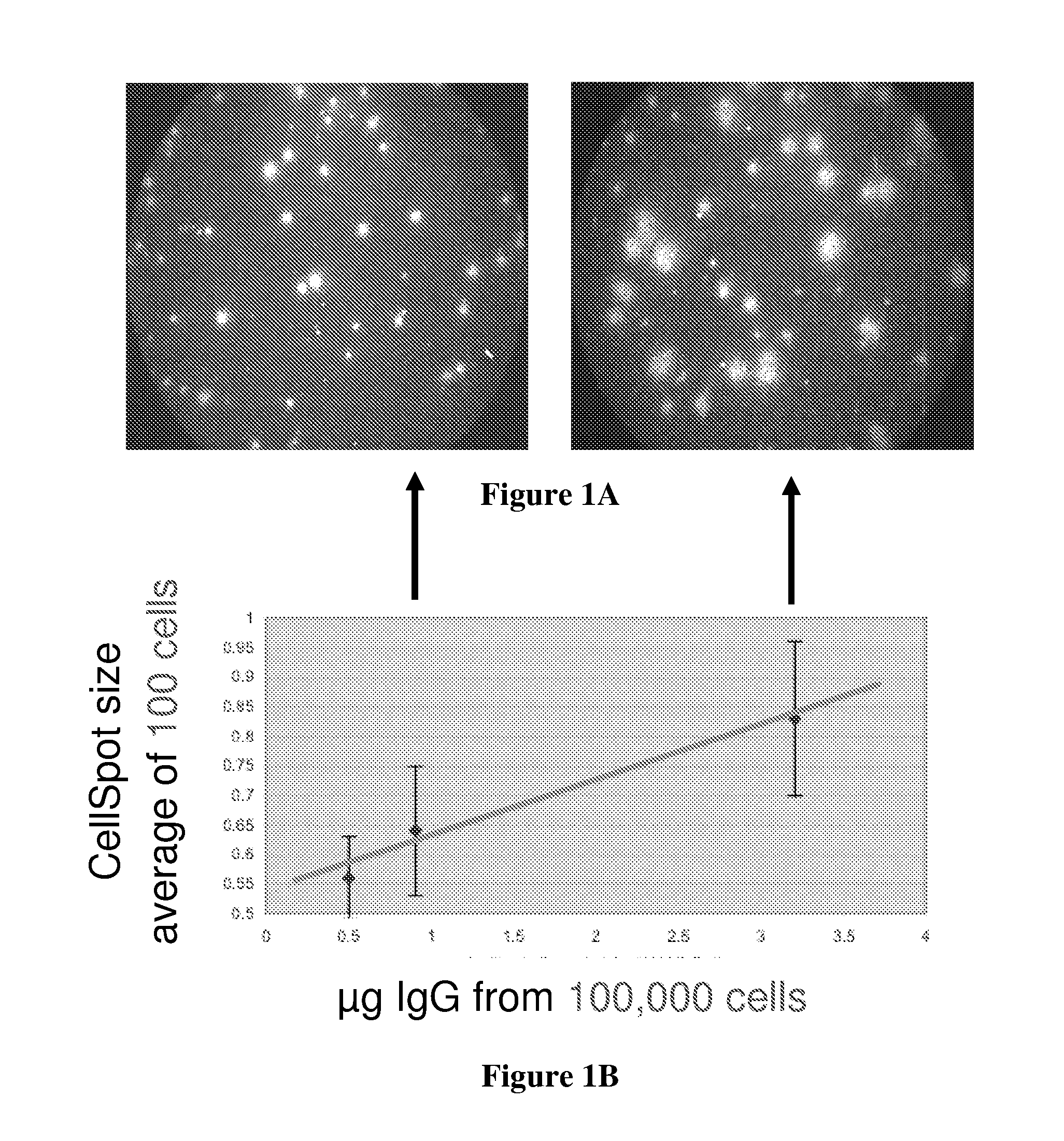
[0094] Hybridoma cells that secrete immunoglobulins were obtained from ATCC and deposited onto a membrane with 0.4 μm pores in contact with an underlying polystyrene surface coated with anti-immunoglobulin. The cells were suspended in 1.2% methylcellulose and to secure the cells, the plate was centrifuged briefly. The secreted IgG passes through the membrane onto the coated polystyrene surface. The membrane containing the cells was supported on a plastic holder that permits it to be removed from the capture surface; the holder was a modified Transwell® material obtained from Costar®, for which a special holder was designed that brings the membrane into contact with the capture surface.

[0095] After incubation for 2 hours, the membrane was removed and the underlying polystyrene surface incubated with detection particles, washed, and then scanned with both a low and high magnification microscope. The results are shown in FIG. 1A which shows the contras...

example 2

Selection of High Secretion Clones

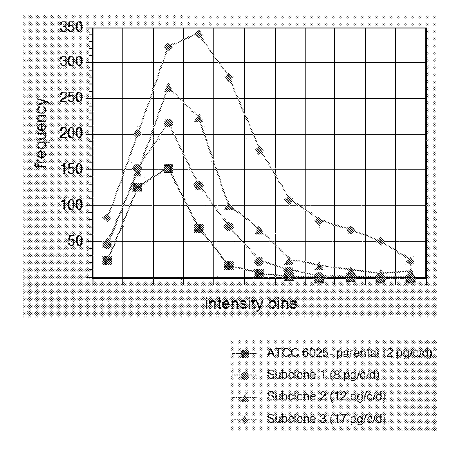
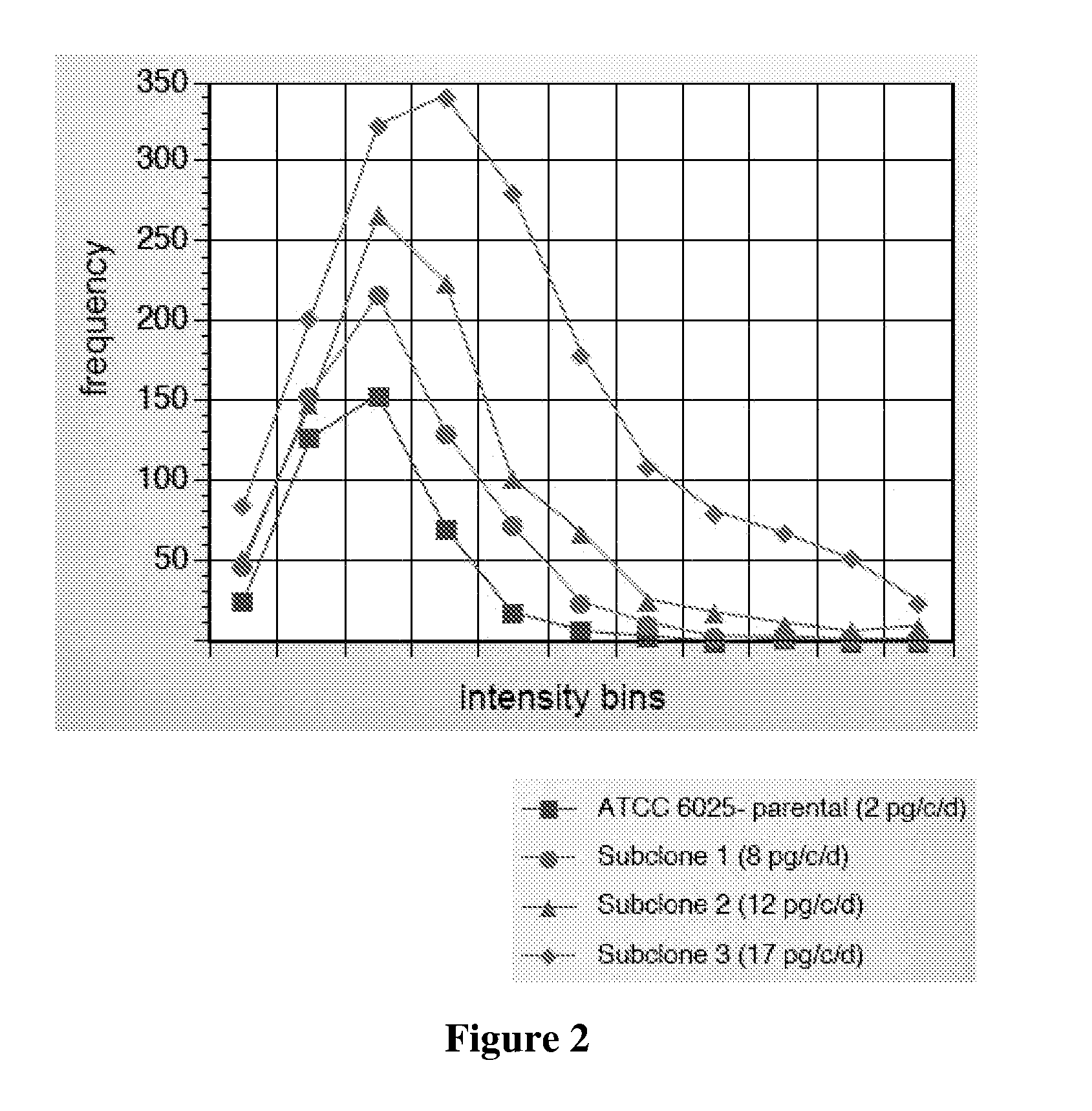
[0097] The cell line ATCC 60525 was separated into 10,000 individual cell assays using the method of Example 1. Three individual cells were picked and cultured as subclones. The subclones were again subjected to the CellSpot™ assay of Example 1 wherein 1,000 cells were assayed for each subclone.

[0098] As shown in FIG. 2, the distribution of secretion levels is shifted to higher secretion levels for the members of the three selected subclone parents resulting in an overall improvement of nine-fold for the highest secretor, as measured by macroscopic supernatant assay.

[0099] The same methodology is applicable to any population of cells that vary in their secretion level, for example a library of transformed CHO cells. Depending on where the DNA for the secreted protein integrates in the genome, expression level will vary. For more reliable identification of high secretors, the cells are allowed to divide in “bins” of 100 parental cells per well of ...

example 3

Determination of Affinity

[0101] Three hybridoma cell lines were determined to secrete antibodies of varying affinity for the same antigen by the Biacore™ commercial instrument method. Each cell line was assayed as set forth in Example 1 using varying concentrations of capture antibody on the capture surface. The clones differed in the frequency of input cells yielding detectable antibodies according to their predetermined affinity as shown in FIG. 3A.

[0102] The assay was conducted by placing a fixed concentration of capture antibody on the surface and counting the number of spots observed at high surface antibody concentration, and assigning a value of 1.00 to that number of spots (100%), as shown on the Y axis of the graph in FIG. 3A. The capture antibody on the surface was then progressively diluted in replicate wells, and the number of spots observed at each dilution. The ratio of this number to that observed at the concentration assigned the value of 1.00 was then plotted on t...


PUM

PropertyMeasurementUnit
Fractionaaaaaaaaaa
Concentrationaaaaaaaaaa
Ratioaaaaaaaaaa

Abstract

A multiplicity of applications of the CellSpot™ assay method are described. Among these applications are extension to integral membrane protein probes, extension to secretion from bacterial cells, identification of antibodies with enhanced affinity, identification of clones with increased secretion levels, and use of massively parallel screening to identify rare efficacious antibodies.

Description

CROSS-REFERENCE TO RELATED APPLICATION [0001] This application claims priority from U.S. provisional application 60 / 830,507 filed 12 Jul. 2006, 60 / 839,174 filed 21 Aug. 2006, 60 / 848,112 filed 29 Sep. 2006, and 60 / 911,483 filed 12 Apr. 2007. The contents of these documents are incorporated herein by reference.TECHNICAL FIELD [0002] The invention concerns methods and compositions related to assays of single cells or multiplexed assays of single or multiple cells where microscopic observation is employed to enhance the efficiency of assays involving secreted proteins. More specifically, the invention is directed to improvements in experimental technique, determination of relative affinity of antibodies, parsing of cell populations for desired features, and application of ELISpot techniques to bacterial systems. BACKGROUND ART [0003] PCT publication WO 2005 / 045396 published 19 May 2005 sets forth the work of the present inventors in adapting conventional ELISpot assays to single cell pr...

Claims


Application Information

Patent Timeline
IPC IPC(8): C12Q1/70C12N5/04C40B20/04G01N33/53C12Q1/68C12N5/07C12N5/071C12N5/078
CPCC07K16/00G01N33/5005G01N33/56966G01N33/6854G01N33/6842G01N33/6845G01N33/56972
Inventor KAUVAR, LAWRENCE M.HARRIMAN, WILLIAM D.COLLARINI, ELLEN J.DUTTA, APRIL
Owner TRELLIS BIOSCIENCE LLC
Learn More