Methods of Enhanced Detection and Therapy of Inflamed Tissues Using Immune Modulation

a technology of immune modulation and enhanced detection, applied in the field of enhanced detection and therapy of inflamed tissues using immune modulation, can solve the problems of heart attack, often death, mortality associated with coronary artery disease, etc., and achieve the effects of improving diagnosis and therapy, increasing diagnostic uptake, and increasing diagnostic selective targeting

Inactive Publication Date: 2008-08-14
THE GENERAL HOSPITAL CORP
View PDF21 Cites 22 Cited by
  • Summary
  • Abstract
  • Description
  • Claims
  • Application Information

AI Technical Summary

Benefits of technology

[0021]It has now been shown that modulators of the immune system can increase the selective targeting of diagnostic and therapeutic compositions to active and vulnerable atheromatous plaques. Methods of the present invention employ immune modulators to increase the uptake of diagnostic or therapeutic compositions by inflammatory cells associated with active atheromatous and vulnerable plaques, thereby improving diagnosis and therapy.
[0036]c) stabilizing, reducing or eliminating the plaque, thereby treating the subject for active atheromatous and vulnerable plaque.

Problems solved by technology

The narrowing of the coronary arteries as a result of the gradual build up of an atherosclerotic plaque deprives the heart muscles of oxygen and results in angina, often death.
However, the major cause of mortality associated with coronary artery disease results not from this gradual narrowing of the blood vessels, but from the sudden rupture of an atherosclerotic plaque.
The formation of a thrombus can rapidly block the flow of blood to the heart muscles, causing a heart attack.
The rupture of atheromatous plaques located in other arteries can also have serious consequences.
For example, the rupture of a plaque in one of the blood vessels supplying the brain can cause a transient ischemic attack or “stroke” and the rupture of a plaque in the peripheral vasculature can block circulation to the limbs or organs, causing serious complications.
However, these procedures do not always prevent the incidence of acute coronary syndrome.
Even with currently available therapeutic approaches, such as lipid lowering, angioplasty and bypass, an unacceptably high incidence of acute coronary syndrome remains (Sacks et al.
Most current methods of plaque detection, several of which are discussed herein, are inadequate for detecting active atheromatous and vulnerable plaques.
Present methods of plaque detection are also inadequate for detecting vulnerable plaques.
Because angioscopic visualization is limited to the surface of the plaque, it is insufficient for use in detecting actively forming atheromatous and / or vulnerable plaques.
However, none has proven to be sufficiently sensitive to identify vulnerable plaques or monitor the formation thereof.
IVUS can detect thin caps and distinguish regions of intermediate density (e.g., intima that is rich in smooth muscle cells and fibrous tissue) from echolucent regions, but current technology does not determine which echolucent regions are composed of cholesterol pools rather than a thrombus, hemorrhage, or some combination thereof.
Moreover, the spatial resolution (i.e., approximately 2 cm) does not distinguish the moderately thinned cap from the high risk cap (i.e., approximately 25-75 μm) and large dense calcium deposits produce acoustic echoes which produce “shadows”, so that deeper plaques can not be imaged.
However, thermography may not provide information about eroded but non-inflamed lesions, vulnerable or otherwise, having a propensity to rupture.
However, long image acquisition time, high costs, limited penetration and a lack of physiologic data render this approach undesirable for detection of actively forming atheromatous and / or vulnerable plaques.
However, it is unlikely that actively forming and / or vulnerable plaques will be reliably differentiated from stable plaques based solely on such lipid:protein ratios.
This means that such methods may not be useful in detecting small, but actively forming or vulnerable plaques.
The usefulness of such methods in detecting small and / or vulnerable plaques is limited by difficulties in distinguishing such plaques from the background of the surrounding tissue.
Although 3D imaging using positron emission tomography (PET) and Single-Photon Computed Tomography (SPECT) is presently in use, the small size of the arteries as compared to the scatter from the large surrounding tissues lowers the utility of these imaging modalities as well.
However, there is generally a relative lack of selectivity of most photosensitizers for atheromatous plaques and more particularly for vulnerable plaques.
Thus, there are deficiencies associated with current methods available for the detection and treatment of atheromatous and vulnerable plaques.
In particular, current diagnostic methods are not able to reliably detect those plaques that are most prone to rupture, and are therefore the most life-threatening.

Method used


Image

  • Methods of Enhanced Detection and Therapy of Inflamed Tissues Using Immune Modulation
  • Methods of Enhanced Detection and Therapy of Inflamed Tissues Using Immune Modulation
  • Methods of Enhanced Detection and Therapy of Inflamed Tissues Using Immune Modulation

Examples


example 1

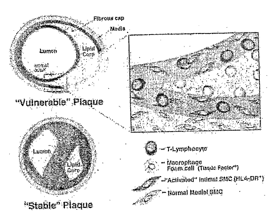
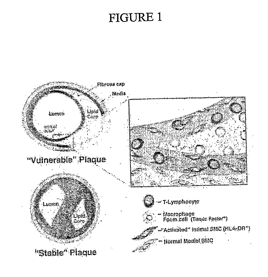
FDG-PET and Intravascular Catheter Imaging in Atherosclerotic Plaques

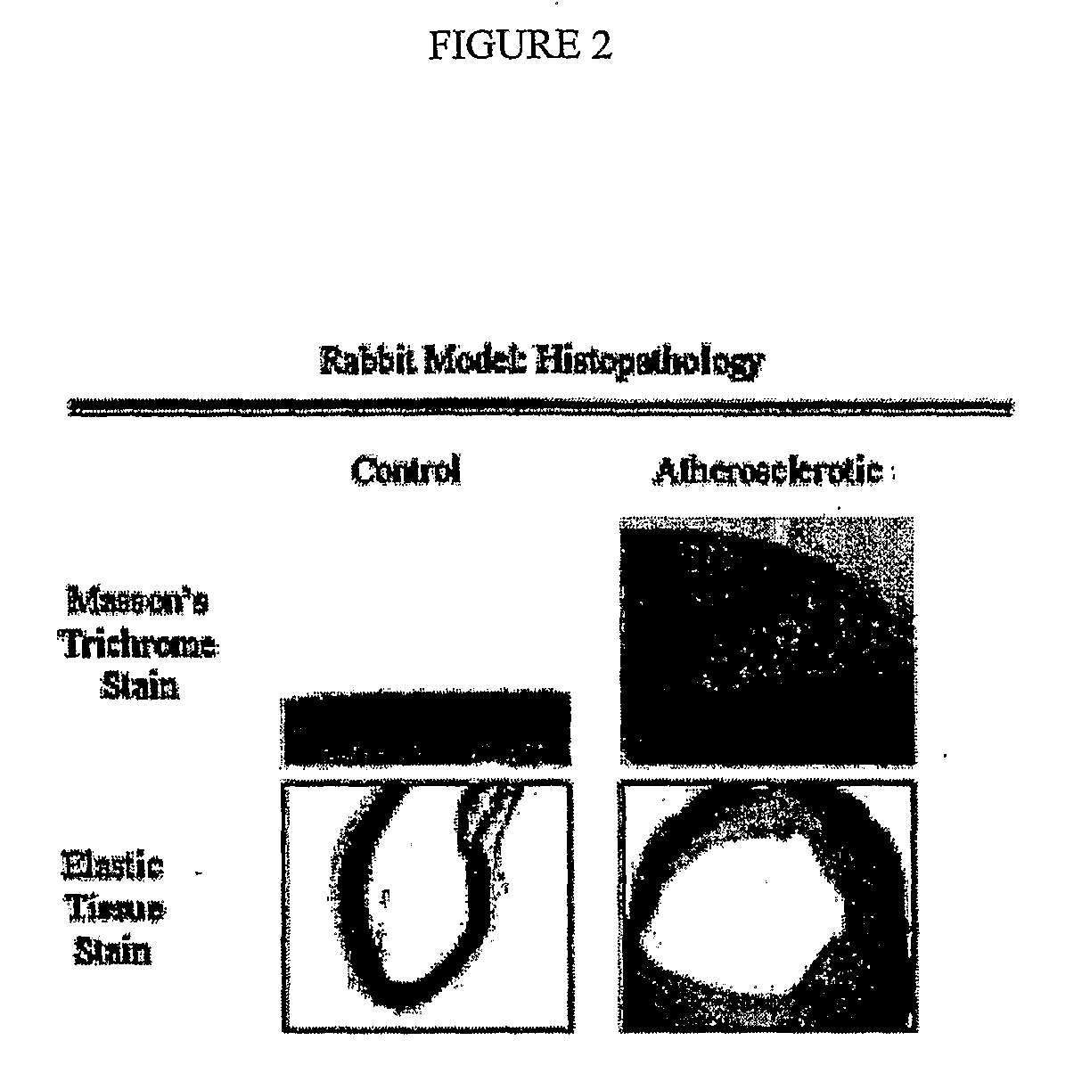
[0201]Male New Zealand white rabbits ranging from 2.5 to 3.0 kilograms in weight (Charles River Breeding Lab) were maintained on a 2% cholesterol-6% peanut oil diet (ICN) for 10 weeks. After 1 week on the peanut oil diet, the abdominal aorta was denuded of endothelium by a modified Baumgartner technique. Briefly, each animal was anesthetized with a mixture of ketamine and xylazine and the right femoral artery was isolated. Subsequently, a 4 F Fogarty embolectomy catheter was introduced via arteriotomy and advanced under fluoroscopic guidance to the level of the diaphragm. The balloon was then inflated to 3 psi (pounds per square inch) above balloon inflation pressure and three passes were made down the abdominal aorta with the inflated catheter. The femoral artery was subsequently ligated and the wound closed. This animal model system is standardly used in the art for the study of active atheromatous and vulnerable...

example 2

Accumulation of 99mTc-Chemotactic Peptide in Rabbit Aorta

[0205]Nuclear methods of targeting inflammatory cells can detect macrophage-rich atherosclerotic lesions. An animal model of atherosclerosis was generated in which macrophage-rich atherosclerotic plaques were induced in New Zealand rabbits by balloon de-endothelialization of the infra-diaphragmatic aorta followed by a high cholesterol diet. At 10 weeks, a 99mtechnetium radiolabeled derivative of bacterial chemotactic peptide CPRA was administered to 7 control rabbits, and to 7 rabbits in which aortic atherosclerotic lesions were induced. This peptide has been shown to bind avidly to leukocytes, with high specificity (Babich, J. W. et al, 1997). At 12 hours after the administration of the radiolabel, the live rabbits were imaged using single photon emission tomography (SPECT). Two investigators that were blinded to the status of the rabbits examined the images. A semi-quantitative scoring system was employed, in which a score o...

example 3

Preparation and Purification of Photosensitizer Compounds

[0206]A photosensitizer composition comprising chlorine6 (“ce6”) coupled to maleylated-albumin) was prepared for targeting to macrophages of a vulnerable plaque animal model system.

[0207]Four photosensitizer compositions were studied (i.e., two BSA-ce6 conjugates and their maleylated counterparts). The N-hydroxy succinimide (NHS) ester of ce6 was prepared by reacting approximately 1.5 equivalents of dicyclohexylcarbodiimide and approximately 1.5 equivalents of NHS with approximately 1 equivalent of ce6 (Porphyrin Products, Logan, Utah) in dry DMSO. After standing in the dark at room temperature for approximately 24 hours, the NHS ester was frozen in aliquots for further use. BSA (Sigma Chemical Co, St Louis, Mo.) (approximately 2×50 mg) was dissolved in NaHCO3 buffer (0.1 M, pH 9.3, approximately 3 ml), and approximately 30 μl and approximately 120 μl of ce6-NHS ester added to respective tubes with vortex mixing. After standin...


PUM

PropertyMeasurementUnit
thickaaaaaaaaaa
thickaaaaaaaaaa
energy tripletaaaaaaaaaa

Abstract

The present invention relates to methods for the detection and therapy of active atheromatous plaques, and in particular vulnerable plaques, whereby immune modulators are used to increase the uptake of diagnostic or therapeutic compositions by the inflammatory cells associated with such plaques.

Description

RELATED APPLICATIONS / PATENTS & INCORPORATION BY REFERENCE[0001]Each of the applications and patents cited in this text, as well as each document or reference cited in each of the applications and patents (including during the prosecution of each issued patent; “application cited documents”), and each of the PCT and foreign applications or patents corresponding to and / or claiming priority from any of these applications and patents, and each of the documents cited or referenced in each of the application cited documents, are hereby expressly incorporated herein by reference. More generally, documents or references are cited in this text, either in a Reference List before the claims, or in the text itself; and, each of these documents or references (“herein-cited references”), as well as each document or reference cited in each of the herein-cited references (including any manufacturer's specifications, instructions, etc.), is hereby expressly incorporated herein by reference.BACKGROUN...

Claims


Application Information

Patent Timeline
Patent Type & Authority Applications(United States)
IPC IPC(8): A61K51/02A61K49/00A61K51/04
CPCA61K41/0057A61K41/0076A61K41/0071A61K51/08
Inventor TAWAKOL, AHMEDHAMBLIN, MICHAEL R.MIGRINO, RAYMOND Q.GELFAND, JEFFREY
Owner THE GENERAL HOSPITAL CORP
Why Eureka
Try Eureka