Intraoperative Imaging Of Hepatobiliary Structures

a technology of intraoperative imaging and biliary organs, which is applied in the field of intraoperative imaging of biliary organs, can solve the problems of erroneously believing, increasing the cost and risk of the procedure for the patient without providing a significant additional, and sometimes complicating the intraoperative assessmen

Inactive Publication Date: 2008-10-09
UNIVERSITY OF ROCHESTER
View PDF10 Cites 12 Cited by
  • Summary
  • Abstract
  • Description
  • Claims
  • Application Information

AI Technical Summary

Problems solved by technology

Unfortunately, common complications of cholecystectomy include injury to the common bile duct, the common hepatic duct, or to the right hepatic duct.
Seventy percent of surgeons regard bile duct injury as an unavoidable complication and there is evidence to suggest that in the majority of duct injuries the surgeon was erroneously convinced he or she correctly identified the damaged duct.
This is a time-consuming process in the operating room, is operator-dependent (requiring a radiologist, a technician, or both), and can produce complications.
Thus, in many cases, the procedure raises costs and risk to the patient without providing a significant additional benefit.
Furthermore, intraoperative assessment is sometimes complicated, needs repeated x-ray imaging and often carries variability in interpretation between observers.
Furthermore, operative cholangiography increases the operative time and is related to a small risk of complications (Voyles, C. R. et al., Ann Surg.
In most of these cases, the surgeon is erroneously satisfied that the biliary structures have been correctly identified and has difficulty interpreting the significance of subsequent events like biliary leakage or jaundice, leading to substantial delay in the diagnosis.
Tumors of the biliary tract are uncommon but serious problems.
Complicated pre and intraoperative imaging of Klatksin tumors, cholangiocarcinomas, is not standardized and is complicated with postprocedural infections or tumor seeding if percutaneous cholangiography is performed, or is inadequate at best when MR or MRCP is used.
These technologies offer deep tissue penetration and high spatial resolution, but are costly and time consuming to implement.

Method used


Image

  • Intraoperative Imaging Of Hepatobiliary Structures
  • Intraoperative Imaging Of Hepatobiliary Structures
  • Intraoperative Imaging Of Hepatobiliary Structures

Examples


example 1

[0048]Intraoperative video angiography is performed with a laser-fluorescence imaging device ((Novadaq Technologies, Inc., Mississauga, Ontario, Canada) consisting of a NIR laser light source and a NIRF-sensitive digital camcorder. For measurements, the unit is positioned 30 to 40 cm from the area of interest. ICG, dissolved in an appropriate carrier, such as saline solution, is then injected as a bolus. The NIR light emitted by the laser light source induces ICG fluorescence. The fluorescence is recorded by a digital video camera, with optical filtering to block ambient and laser light so that only ICG fluorescence is captured. Images can be observed on screen in real time (at approximately 25 to 30 images / sec (PAL or NTSC)). The images can be reviewed and stored on the digital video camera or transferred to a computer or to storage media.

example 2

[0049]In an initial clinical trial, fifteen (15) subjects are enrolled and assessed during the conduct of the trial. All study subjects receive standard of care assessments for their pre- and post-operative care. Candidates for this study meet all of the following inclusion criteria, and do not meet any of the listed exclusion criteria. The inclusion criteria are: subject (or legal representative) is willing and able to provide an informed consent; subject is willing and able to comply with the study procedures, a urine pregnancy test for women of reproductive age prior to surgery shows the woman is not pregnant, and the subject is scheduled for biliary surgery. The exclusion criteria are: subject has significant liver disease, cirrhosis or liver insufficiency with abnormal liver function tests, as total bilirubin>1.5×normal and / or SGOT>2×normal, subject has uremia, serum creatinine>2.5 mg / dl, subject has a previous history of adverse reaction or allergy to ICG, iodine, shellfish or...

example 3

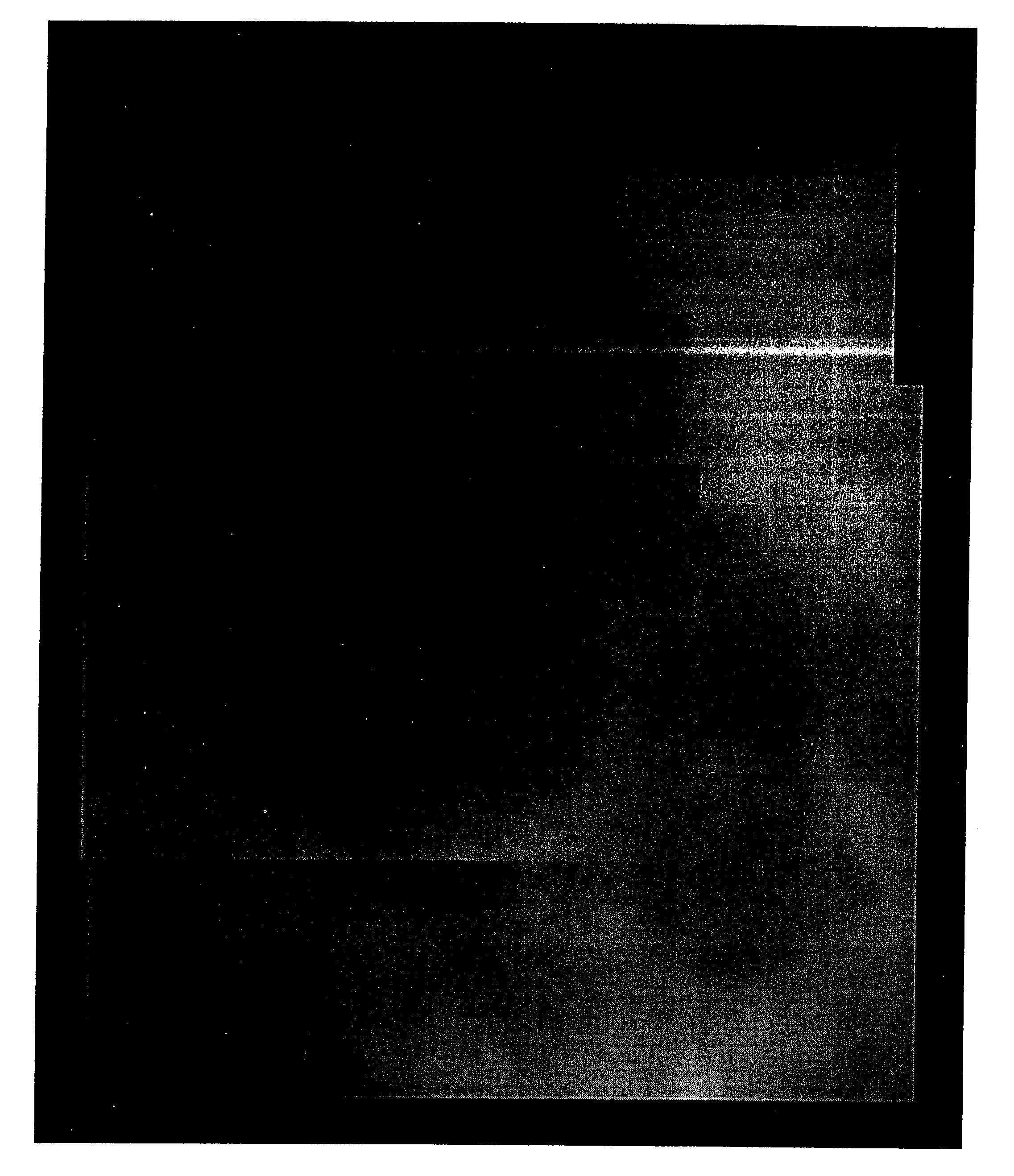
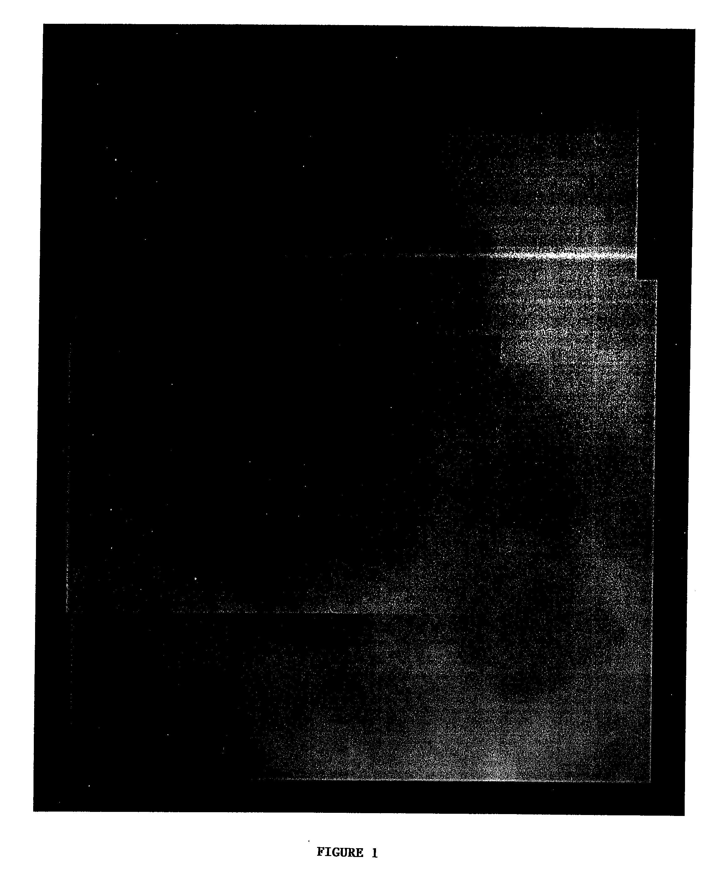
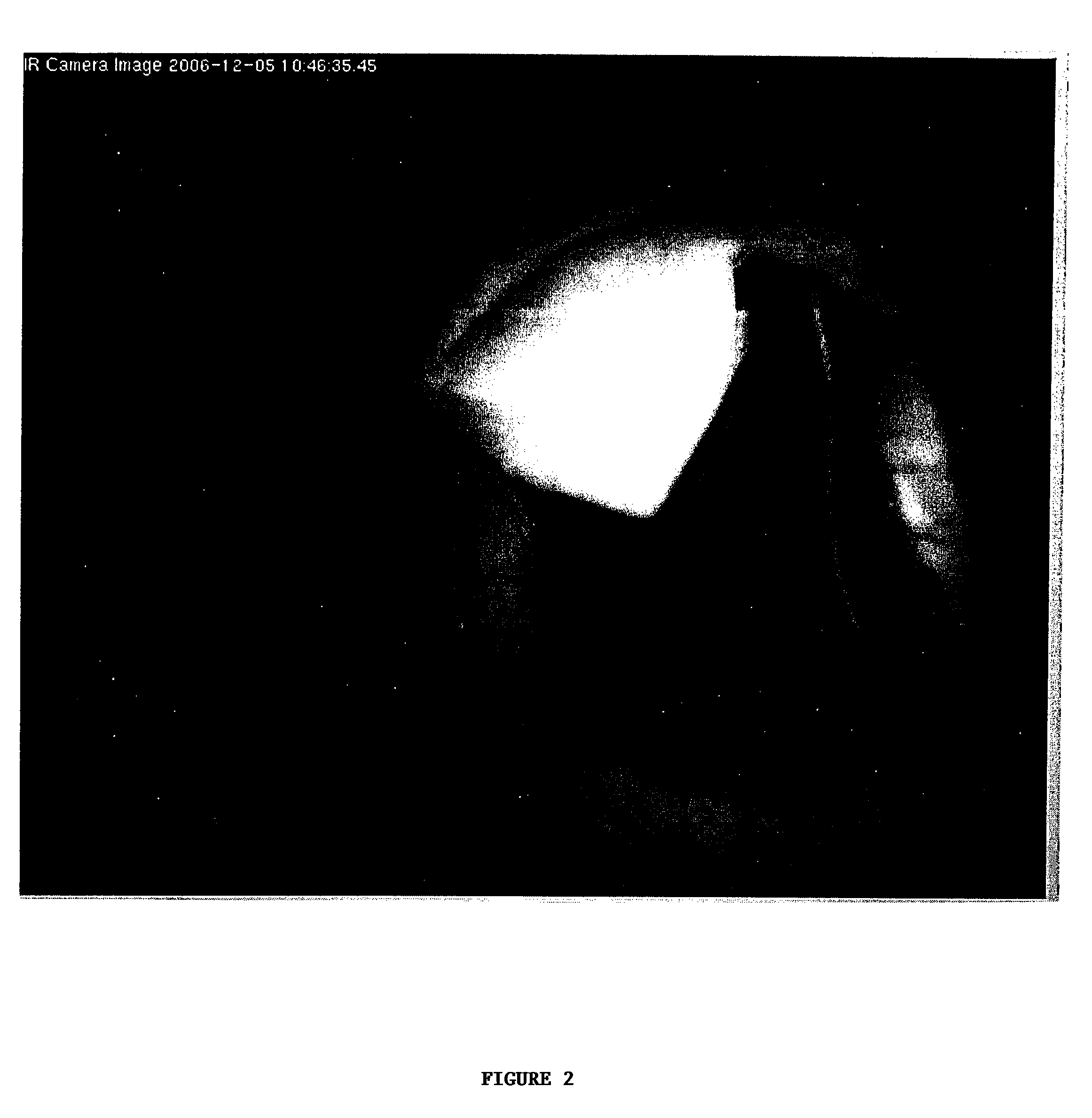
[0053]This Example reports the results of an investigation of liver visualization by fluorescent dye in an adult pig model. An adult pig (approximately 50-60 kilograms) was placed under appropriate anesthesia and its liver subjected to radiofrequency (RF) emissions from a RF needle electrode. The liver looked normal upon visual examination under normal light. ICG was administered intravenously as a bolus injection and the liver was visualized by fluorescent illumination using a SPY™ imaging system (Novadaq Technologies, Inc., Mississauga, Ontario, Canada). FIG. 1 is a composite of photographs taken of the light emitted from the liver under laser excitation light illumination. The lighter areas to the right and bottom sides of the photos indicate normal liver tissue. The darker areas in the middle shows areas of low perfusion, while the black area on the left side of the photos had no visible perfusion and was likely dead. Upon palpation, the light areas were flexible and normal, whi...


PUM


Abstract

The invention provides methods for visualizing hepatobiliary structures, or lesions in a liver, intraoperatively by use of fluorescent dyes.

Description

CROSS-REFERENCES TO RELATED APPLICATIONS[0001]This application claims the benefit of U.S. Provisional Application No. 60 / 850,041, filed Oct. 6, 2006, the contents of which are incorporated herein by reference.STATEMENT AS TO RIGHTS TO INVENTIONS MADE UNDER FEDERALLY SPONSORED RESEARCH AND DEVELOPMENT[0002]Not applicable.REFERENCE TO A “SEQUENCE LISTING,” A TABLE, OR A COMPUTER PROGRAM LISTING APPENDIX SUBMITTED ON A COMPACT DISK.[0003]Not applicableBACKGROUND OF THE INVENTION[0004]Laparoscopic cholecystectomy (removal of the gall bladder) represents the most common gastrointestinal surgical procedure, with some 700,000 operations annually performed in the United States. The laparoscopic approach has become the treatment of choice for symptomatic cholelithiasis because of its advantages compared to the classic open technique, in terms of shorter hospital stay, reduced postoperative pain and quicker return to normal daily activities (Deziel et al., Am J Surg, 165:9-14 (1993); Hannan e...

Claims


Application Information

Patent Timeline
Patent Type & Authority Applications(United States)
IPC IPC(8): A61B5/00
CPCA61K49/0034
Inventor GOLIJANIN, DRAGANWOOD, RONALDREEDER, JAY E.
Owner UNIVERSITY OF ROCHESTER
Why Eureka
Try Eureka