Aptamer-based methods for identifying cellular biomarkers

a technology of cellular biomarkers and aptamer-based methods, which is applied in the direction of fluid pressure measurement, liquid/fluent solid measurement, peptides, etc., can solve the problems of insufficient number of samples, limited number of molecules identified as effective tumor markers, and non-standard technology platforms. achieve the effect of easy immobilization

Inactive Publication Date: 2009-05-07
TAN WEIHONG +1
View PDF8 Cites 67 Cited by
  • Summary
  • Abstract
  • Description
  • Claims
  • Application Information

AI Technical Summary

Benefits of technology

[0017]The subject invention provides methods for identifying cell biomarkers using highly specific and / or selective oligonucleotide ligands. The subject invention also discloses methods for producing such oligonucleotide ligands for use in identifying cell biomarkers. In certain related embodiments, the ligands are derived from an in vitro evolution process called SELEX (Systematic evolution of ligands by exponential enrichment) and can be synthesized in large scale by DNA synthesizer easily. Biomarkers of interest are subsequently captured in cell lysate by these oligonucleotide ligands, which are bound to a biotin-tag, and the resultant complexes are easily immobilized on a magnetic solid support using streptavidin coated magnetic beads.
[0019]In related embodiments, DNA aptamers are developed directly from tumor cells to function as highly specific and selective probes. These probes are tagged with biotin and subsequently bound to surface targets of leukemia cells to facilitate the discovery of potential new biomarkers via magnetic strategies. In certain embodiments, aptamers highly specific for a T-cell acute lymphoblastic leukemia (T-ALL) cell line are selected in accordance with the subject invention. Such T-ALL-aptamers have high affinity and excellent specificity. These molecular probes were utilized with magnetic strategies (biotin-labeling, streptavidin coated magnetic beads, and magnetic support stand) to capture and purify cancer cell membrane targets for the identification of disease biomarkers. Using the disclosed methods, a trans-membrane receptor protein tyrosine kinase 7 (PTK7) was identified to be the target of a T-ALL specific aptamer. The finding of high expression of PTK7 on T-ALL cells and the simultaneous development of an excellent aptamer probe for it assists in practical and reliable diagnosis of related leukemia.
[0021]In a related embodiment, the present invention provides a cell-based strategy (cell-SELEX) that generates a group of aptamers (designer DNA / RNA probes) that can specifically recognize an individual cancer cell type, without having prior knowledge about the cancer biomarker. These probes are then utilized to facilitate extraction, purification, and identification of the membrane biomarkers on the cancer cells. The identification of the membrane targets is realized through magnetized collection and separation and analysis via routine affinity chromatography coupled with mass spectrometry. Upon discovery of the cancer-specific biomarkers of the invention, aptamer probes (or antibodies) are generated for the recognition of these biomarkers for diagnosis of the corresponding disease, which greatly expedites the clinical application of the newly discovered biomarkers.

Problems solved by technology

Despite the enormous research efforts in the development of cancer biomarkers, a very limited number of molecules have been identified as effective markers for tumors.
The main reasons behind this are likely insufficient number of samples and non-standardized technology platforms.
It is noteworthy that while these methods have been used for biomarker discovery for some time, the number of biomarkers identified and widely accepted is still very limited.
In fact, systematic production of a panel of antibodies for molecular differentiation of cancer cells is very difficult.
This is believed to be due to subtle differences among patients that are not detectable using current technologies.
However, identification of molecular fingerprints of cancers remains extremely challenging, not to mention the development of corresponding molecular probes.
However, these morphologic features are difficult to be used to carry out early cancer diagnosis, or to evaluate the complex molecular alterations that lead to cancer progression (Luo, J., Isaacs, W. B., Trent, J. M. & Duggan, D. J. (2003) Cancer Invest.
Nonetheless, identification of molecular signatures of a particular cancer remains a great challenge if not impossible, which is reflected by the fact that very few biomarkers are available for effective cancer diagnosis.
However, these antigens are usually expressed on both neoplastic cells and normal hematopoietic cells, and could not accurately reflect the molecular features of the cancer cells.
In fact, no panels of monoclonal antibodies are available to reliably distinguish tumor cells from their normal counterparts.
This is due to the technical difficulties in systematic development of antibodies for unknown surface biomarkers.
However, identifying molecular differences between any two types of cells is not an easy task with current technologies.
For example, discovery of unknown molecular features of diseased cells using molecular probes is almost impractical because, most of today's methodologies rely on known biomarkers for the development of corresponding molecular probes, which has been proved insufficient for addressing many emerging medical problems.
Currently the application of aptamers towards medical research and application is limited due to the lack of aptamers for systems of medical relevance.

Method used


Image

  • Aptamer-based methods for identifying cellular biomarkers
  • Aptamer-based methods for identifying cellular biomarkers
  • Aptamer-based methods for identifying cellular biomarkers

Examples


example 1

Materials

Cell Lines and Reagents

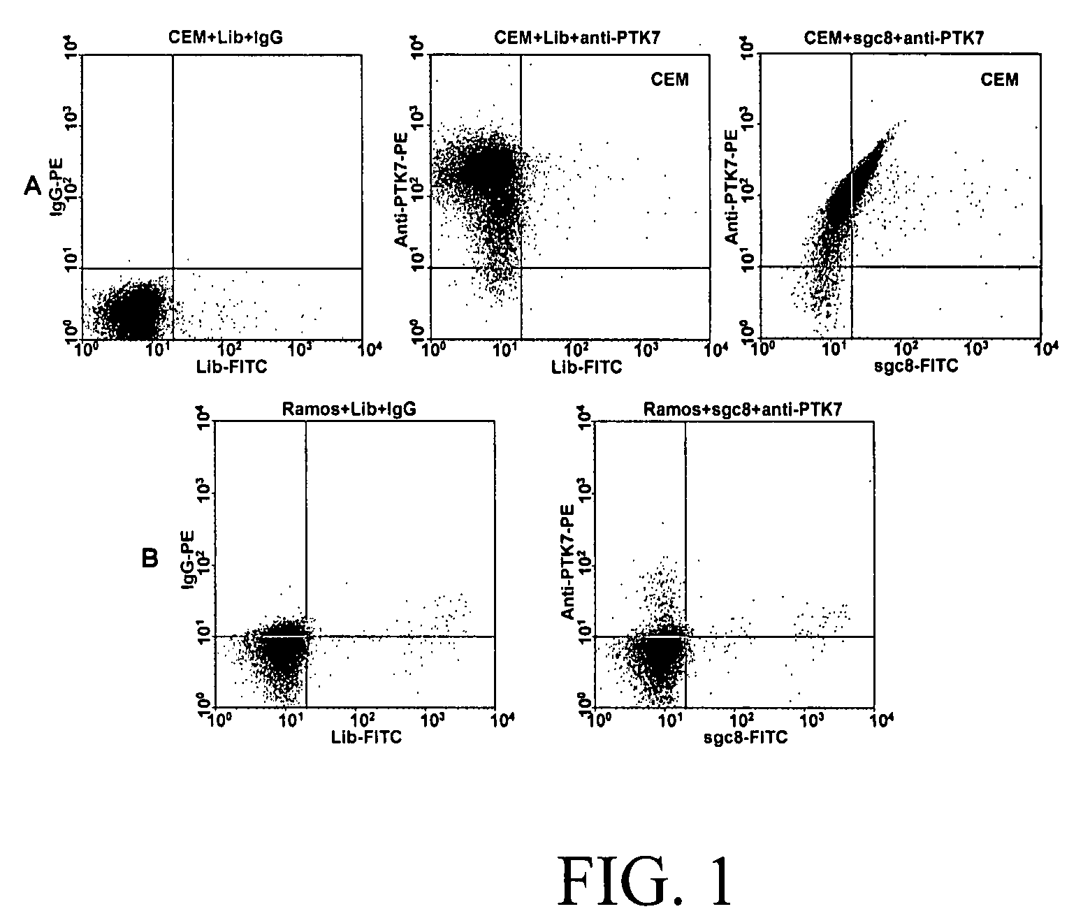
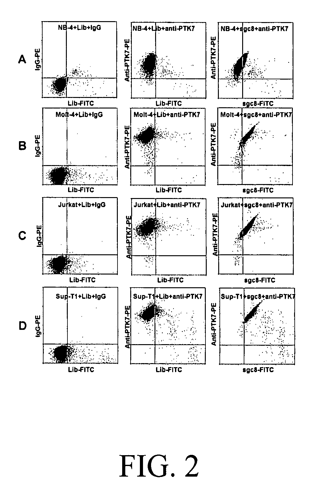
[0055]CCRF-CEM (CCL-119, T-cell lines, human acute lymphoblastic leukemia), Ramos, (CRL-1596, B-cell line, human Burkitt's lymphoma), Toledo (CRL-2631, B-cell line, human diffuse large cell lymphoma), Sup-T1 (CRL-1942, T-cell lines, human lymphoblastic leukemia), Jurkat (TIB-152, human acute T cell leukemia), Molt-4 (CRL-1582, T-cell lines, human acute lymphoblastic leukemia), were obtained from ATCC (American Type Culture Collection). NB-4 (acute promyelocytic leukemia) was obtained from the Department of Pathology, University of Florida). All the cells were cultured in RPMI 1640 medium (ATCC) supplemented with 10% fetal bovine serum (FBS) (heat inactivated, GIBCO) and 100 IU / mL penicillin-Streptomycin (Cellgro). Cells were washed before and after incubation with wash buffer (4.5 g / L glucose and 5 mM MgCl2 in Dulbecco's phosphate buffered saline with calcium chloride and magnesium chloride (Sigma)). Binding buffer used for selection was prepared by a...

example 2

Cell-SELEX

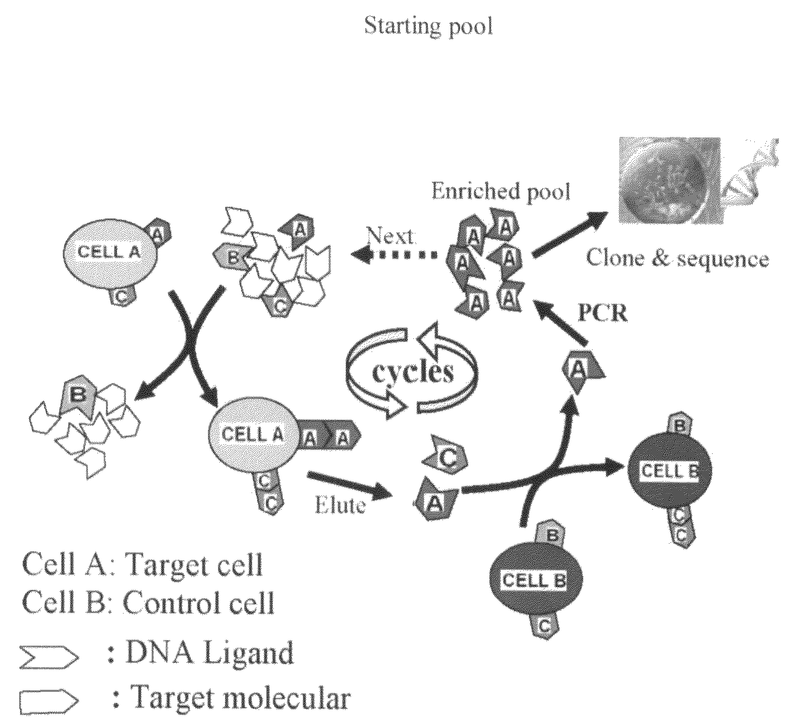
[0082]Cell-based SELEX procedure (Aptamer selection): The cell-SELEX procedure is schematically shown in FIG. 6. Synthesized ssDNA library was incubated with target cells. After washing, the bound DNAs were eluted by heating. The collected DNAs were then incubated with negative cells for counter-selection in order to remove the sequences binding to coexisting molecules on both cells. After centrifugation, the supernatant was collected and the selected DNA pool was amplified by PCR. The PCR products were separated into ssDNAs for next round selection. After the DNA pool reached certain cell-binding affinity, the enriched pool was cloned and sequenced. Aptamers were identified from the sequenced pool. These aptamers have Kd in the range of nM to pM: sgc3: 1.97 nM, sgc4: 26.6 nM, sgc8: 0.8 nM, sgd2: 7.2 nM, sgd3: 3.58 nm, and sgd5: 70.8 nM. Aptamer sgd5 was selected from Toledo cells, a human diffuse large B cell lymphoma cell line. All the other aptamers were from CCRF-CEM c...

example 3

Cell Lines and Buffers

[0101]CCRF-CEM (CCL-119, T-cell lines, human acute lymphoblastic leukemia), Ramos, (CRL-1596, B-cell line, human Burkitt's lymphoma), Toledo (CRL-2631, B-cell line, human diffuse large cell lymphoma), Sup-T1(CRL-1942, T-cell lines, human lymphoblastic leukemia), Jurkat (TIB-152, human acute T cell leukemia), Molt-4 (CRL-1582, T-cell lines, human acute lymphoblastic leukemia), SUP-B15 (CRL-1929, B-lymphoblast, human acute lymphoblastic leukemia) and U266 (TIB-196, B-lymphocyte, human myeloma, plasmacytoma) were obtained from ATCC (American Type Culture collection). Mo2058 (Mantle-cell lymphoma, Epstein-Barr Virus-positive cell line) and NB-4 (acute promyelocytic leukemia) were obtained from Department of Pathology, University of Florida). All the cells were cultured in RPMI 1640 medium (ATCC) supplemented with 10% fetal bovine serum (FBS) (heat inactivated, GIBCO) and 100 IU / mL penicillin-Streptomycin (Cellgro). Cells were washed before and after incubation with...


PUM

PropertyMeasurementUnit
pHaaaaaaaaaa
volumeaaaaaaaaaa
affinityaaaaaaaaaa

Abstract

In this invention, a biomarker discovery method has been developed using specific biotin-labeled oligonucleotide ligands and magnetic streptavidin beads. In one embodiment, the oligonucleotide ligands are firstly generated by whole-cell based SELEX technique. Such ligands can recognize target cells with high affinity and specificity and can distinguish cells that are closely related to target cells even in patient samples. The targets of these oligonucleotide ligands are significant biomarkers for certain cells. These important biomarkers can be captured by forming complexes with biotin-labeled oligonucleotide ligands and collecting the complexes using magnetic streptavidin beads, whereupon the captured biomarkers are analyzed to identify the biomarkers. Analysis of biomarkers include HPLC-Mass Spectroscopy analysis, polyacrylamide gel electrophoresis, flow cytometry, and the like. The identified biomarkers can be used for pathological diagnosis and therapeutic applications. Using the disclosed methods, highly specific biomarkers of any kinds of cells, in particular cancer cells, can easily be identified without prior knowledge of the existence of such biomarkers.

Description

CROSS-REFERENCE TO A RELATED APPLICATION[0001]This application claims the benefit of U.S. provisional application Ser. No. 60 / 831,749, filed on Jul. 17, 2006, which is hereby incorporated by reference in its entirety, including all figures and tables.GOVERNMENT SUPPORT[0002]The subject matter of this application has been supported in part by U.S. Government Support under NIH GM66137 and NSF EF0304569. Accordingly, the U.S. Government has certain rights in this invention.BACKGROUND OF THE INVENTION[0003]Molecular biomarkers are crucial for diagnosis of diseases and for predicting disease development. Potentially, some biomarkers can also be the targets of therapeutic agents for tumor-specific drug delivery in cancer treatments. Depending on the location, cancer markers can be present in the serum, such as the classic prostate-specific antigen (PSA) for prostate cancer (M. J. Barry, N. Engl. J. Med. 344, 1373 (2001)), or expressed directly on cells, such as the Her-2 receptor in breas...

Claims


Application Information

Patent Timeline
Patent Type & Authority Applications(United States)
IPC IPC(8): C12Q1/68G01N33/566B01D59/44
CPCC12N15/111C12N2320/11C12N2310/16C12N15/115
Inventor TAN, WEIHONGSHANGGUAN, DIHUA
Owner TAN WEIHONG
Why Eureka
Try Eureka